Vertebrobasilar Artery
MYO CORE
Core Anatomy
The vertebrobasilar system is the principal arterial network of the posterior circulation, formed by the vertebral arteries and basilar artery. It supplies the brainstem, cerebellum, thalamus, occipital lobes, and upper cervical spinal cord, supporting multiple neural centers essential for life and neurological function.
OVERVIEW
The vascular territory supplied by the vertebrobasilar system includes the medulla oblongata, pons, midbrain, cerebellum, occipital lobes, inferomedial temporal lobes, posterior thalamus, inner ear, and upper cervical spinal cord.
These regions contain numerous nuclei, tracts, and integrative centers responsible for consciousness, respiration, cardiovascular regulation, ocular movements, hearing, balance, coordination, sensorimotor integration, and visual processing.
Because the posterior circulation supplies many of the most vital structures in the nervous system, vertebrobasilar compromise frequently produces severe neurological deficits that are often disproportionate to the size of the vascular lesion.
Even relatively small ischemic insults may disrupt essential brainstem functions, resulting in cranial nerve dysfunction, respiratory instability, impaired consciousness, or catastrophic neurological deterioration.
Consequently, the vertebrobasilar system is considered one of the most clinically significant vascular networks within the human body.
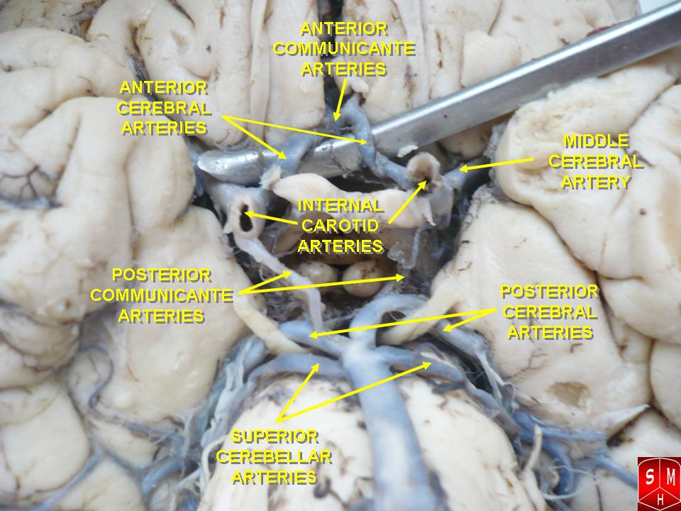
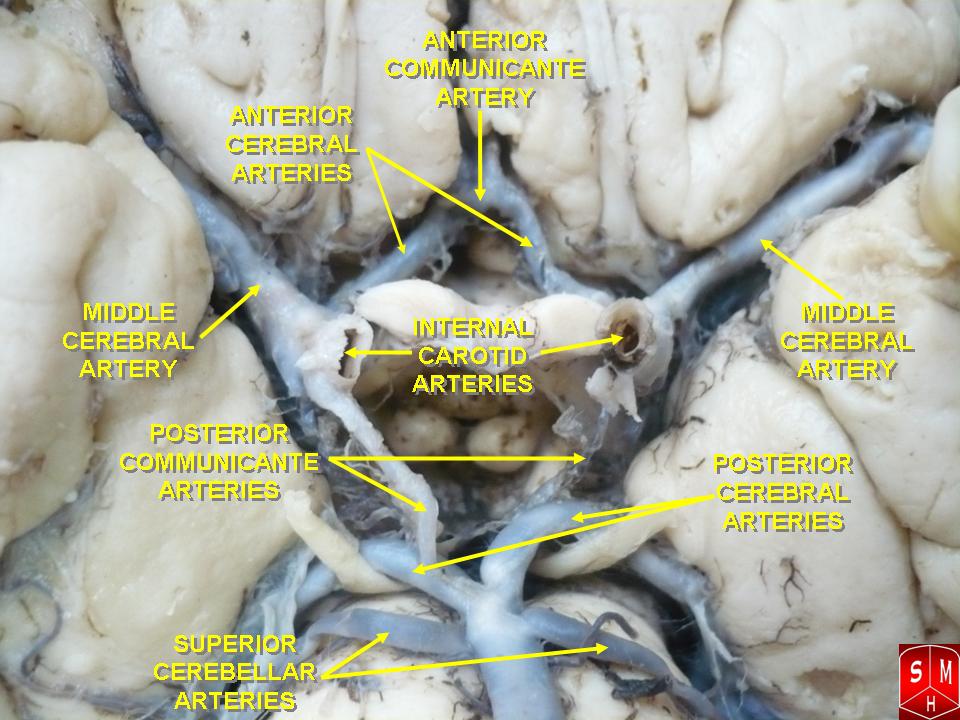
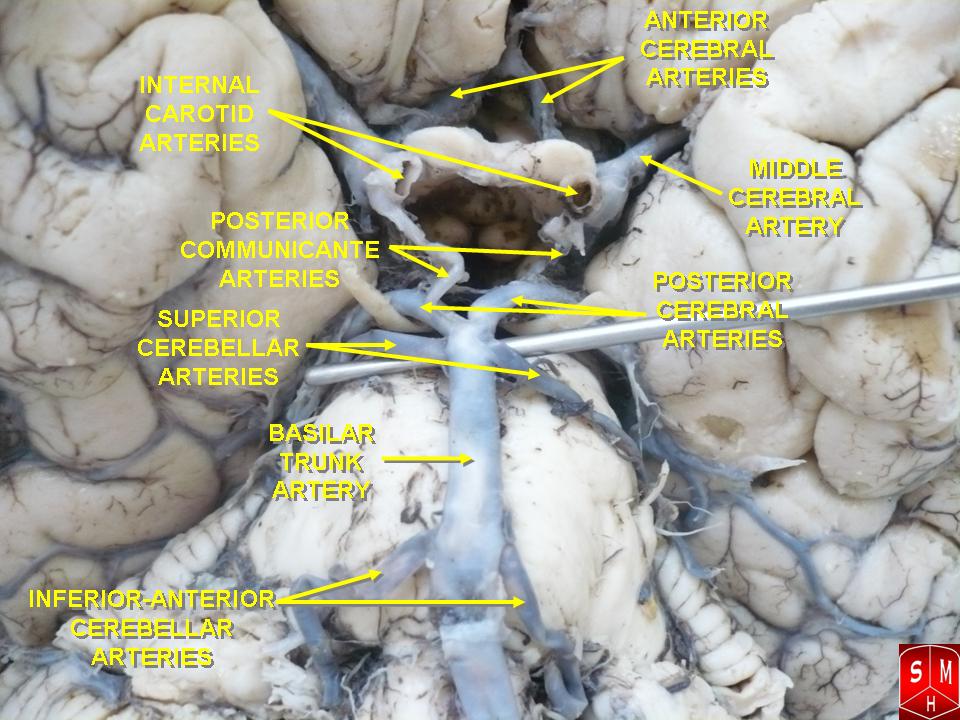
ANATOMY
Formation
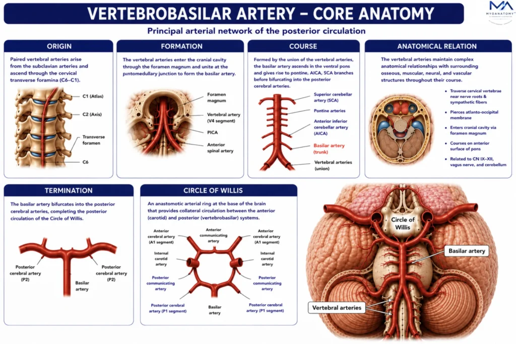
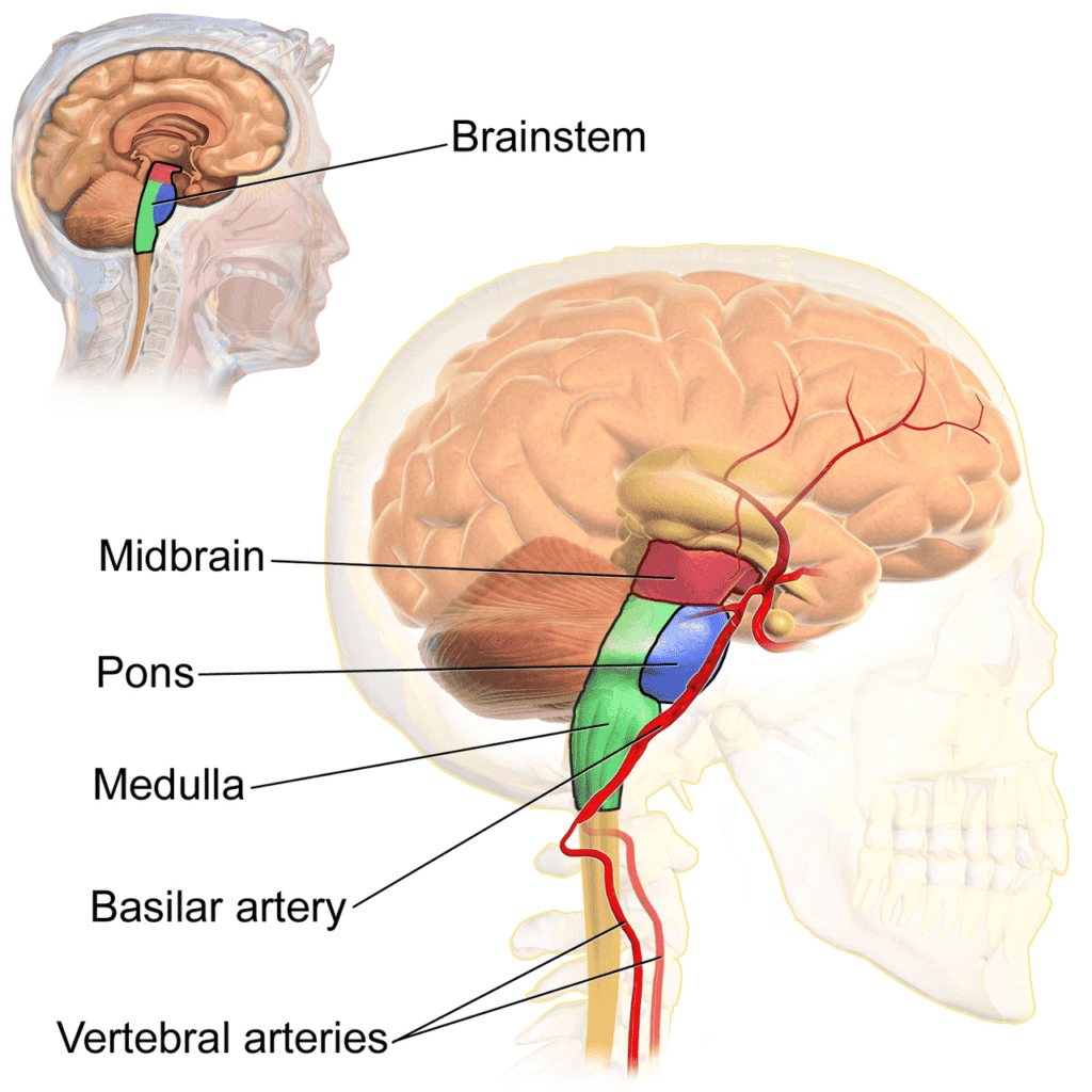
The vertebrobasilar system originates from the right and left vertebral arteries, which usually arise as the first branches of their respective subclavian arteries. Each vertebral artery ascends through the cervical region before entering the cranial cavity through the foramen magnum. After traversing the posterior cranial fossa, the two vessels converge at the pontomedullary junction along the ventral aspect of the brainstem.
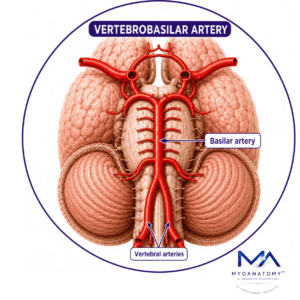
Fusion of the paired vertebral arteries forms the basilar artery, a single midline vessel that ascends within the prepontine cistern. This union establishes a continuous arterial conduit connecting the extracranial circulation with the posterior cerebral circulation.
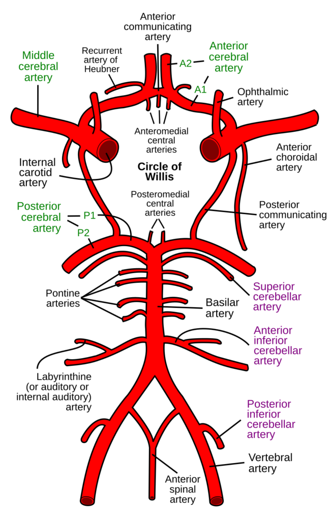
Before their convergence, the vertebral arteries contribute substantially to posterior circulation perfusion through numerous branches supplying the cervical spinal cord, medulla oblongata, meninges, and cerebellum. Among these branches, the posterior inferior cerebellar artery (PICA) is particularly important because it supplies extensive portions of the lateral medulla and inferior cerebellum.
The formation of the basilar artery is the integration of two major vascular channels into a single arterial trunk responsible for maintaining perfusion of the brainstem and posterior cerebrum. Pathology affecting either vertebral artery may consequently influence perfusion throughout the entire vertebrobasilar system.
Exam Question
Describe the formation of the vertebrobasilar system, including the origin, convergence, and major vascular contributions of the vertebral arteries.
Course
After arising from the subclavian arteries, the vertebral arteries ascend through the transverse foramina of the cervical vertebrae, typically beginning at the level of C6. They continue superiorly through the cervical spine before curving posteriorly around the lateral mass of the atlas, where they occupy a vulnerable position within the suboccipital region.
The arteries then pierce the atlanto-occipital membrane and dura mater to enter the cranial cavity through the foramen magnum. Within the posterior cranial fossa, each vertebral artery ascends along the anterolateral surface of the medulla oblongata, giving rise to spinal, cerebellar, and perforating branches.
At the pontomedullary junction, the vertebral arteries unite to form the basilar artery. The basilar artery subsequently ascends within the basilar groove located on the ventral surface of the pons, remaining closely applied to the brainstem throughout its course.
Along its trajectory, the basilar artery gives rise to numerous perforating pontine arteries, the anterior inferior cerebellar arteries (AICA), labyrinthine arteries, and superior cerebellar arteries (SCA). At the superior border of the pons, adjacent to the pontomesencephalic junction, it terminates by dividing into the right and left posterior cerebral arteries.
This uninterrupted vertebral-basilar-posterior cerebral pathway forms the central arterial axis of the posterior circulation and constitutes the primary route through which blood reaches the brainstem, cerebellum, and posterior cerebrum.
Exam Question
Trace the course of the vertebral and basilar arteries from their cervical origin to their intracranial termination, emphasizing key anatomical landmarks and branches.
Anatomical Relation
The vertebral arteries maintain complex anatomical relationships with surrounding osseous, muscular, neural, and vascular structures throughout their course.
Within the neck they traverse the transverse foramina of the cervical vertebrae and lie in close proximity to cervical nerve roots, sympathetic fibers, deep cervical muscles, and the atlantoaxial and atlanto-occipital joints.
As they enter the cranial cavity, the vertebral arteries ascend along the lateral surfaces of the medulla oblongata, where they are intimately related to the pyramids, inferior olivary nuclei, inferior cerebellar peduncles, and lower cranial nerves. The glossopharyngeal nerve (CN IX), vagus nerve (CN X), accessory nerve (CN XI), and hypoglossal nerve (CN XII) all maintain close spatial relationships with vertebral artery branches and perforators.
The basilar artery occupies the basilar groove on the anterior surface of the pons, lying immediately anterior to pontine nuclei, corticospinal tracts, ascending sensory pathways, reticular formation structures, and numerous cranial nerve nuclei. Small perforating arteries arise directly from the posterior surface of the basilar artery and penetrate the pons to supply these critical neural structures.
Superiorly, the basilar artery is related to the midbrain, interpeduncular cistern, oculomotor nerves (CN III), posterior cerebral arteries, superior cerebellar arteries, and posterior communicating arteries.
Exam Question
Analyze the anatomical relationships of the vertebral and basilar arteries with adjacent cranial nerves, brainstem structures, and surrounding neurovascular components.
Termination
The basilar artery terminates at the superior border of the pons by bifurcating into the right and left posterior cerebral arteries (PCAs), which constitute the terminal branches of the vertebrobasilar system.
Following their origin, the posterior cerebral arteries course around the cerebral peduncles and distribute blood to extensive posterior cerebral territories. Their vascular distribution includes the occipital cortex, inferomedial temporal lobe, splenium of the corpus callosum, posterior thalamus, portions of the midbrain, and associated deep white matter pathways.
Through these terminal branches, the vertebrobasilar system becomes the dominant arterial supplier of the visual cortex and major components of the posterior cerebrum. Consequently, lesions affecting the distal basilar artery or proximal posterior cerebral arteries frequently produce visual field deficits, cortical blindness, thalamic syndromes, memory disturbances, and complex neurocognitive impairments.
The basilar bifurcation therefore represents the anatomical and functional transition between the brainstem circulation and the posterior cerebral circulation.
Exam Question
Discuss the termination of the basilar artery and evaluate the functional importance of the posterior cerebral arteries in supplying the posterior cerebrum.
Circle of Willis
The vertebrobasilar system forms the posterior component of the Circle of Willis. Each posterior cerebral artery communicates with the internal carotid circulation through a posterior communicating artery, creating a collateral link between the anterior and posterior cerebral circulations.
Although flow through these channels is usually limited under normal conditions, they become essential during arterial stenosis or occlusion, allowing redistribution of blood to preserve cerebral perfusion. The effectiveness of this collateral network depends on the anatomical completeness of the Circle of Willis, which varies considerably among individuals and influences susceptibility to ischemic injury.
Exam Question
Assess the role of the vertebrobasilar system in the Circle of Willis and explain how collateral circulation may preserve cerebral perfusion during arterial compromise.
SUMMARY TABLE