Internal Carotid Artery
MYO CORE
Core Anatomy
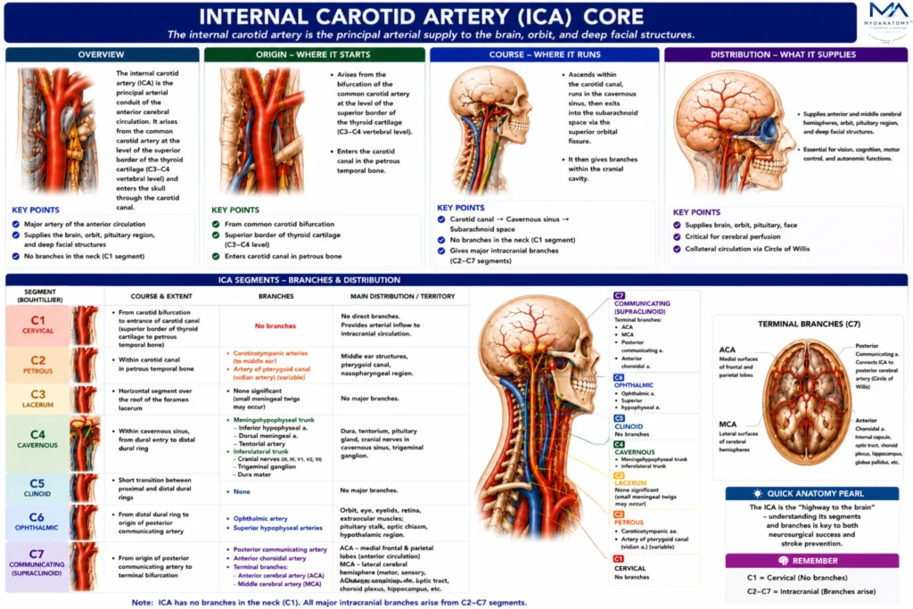
The internal carotid artery is the principal arterial conduit of the anterior cerebral circulation, providing a direct high-capacity vascular pathway between the systemic circulation and the brain. Through its extensive intracranial branches, it supplies the cerebral hemispheres, visual apparatus, pituitary region, and deep cerebral structures essential for higher neurological function.
OVERVIEW
Unlike the external carotid artery, which distributes blood to numerous extracranial tissues through multiple cervical branches, the Internal Carotid Artery ( ICA) is specialized for uninterrupted intracranial perfusion and therefore gives no branches in the neck.
This unique anatomical characteristic reflects its primary role in maintaining continuous delivery of oxygen and glucose to neural tissues with exceptionally high metabolic demands.
From its origin at the carotid bifurcation, the ICA traverses multiple anatomical environments – including the cervical compartment, petrous temporal bone, cavernous sinus, and subarachnoid space – before terminating as the major arteries of the forebrain. Along this course, it becomes intimately associated with the optic apparatus, pituitary gland, cavernous sinus, autonomic plexuses, and several cranial nerves.
Through its terminal branches and communicating arteries, the ICA forms a major component of the Circle of Willis, providing both direct cerebral perfusion and collateral vascular protection.
Consequently, the ICA occupies a central position in cerebrovascular physiology, neuroanatomy, neuroradiology, vascular surgery, and stroke medicine.
ANATOMY
Origin
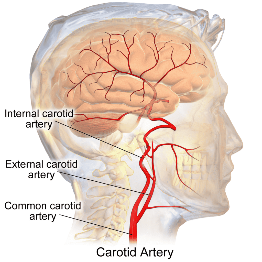
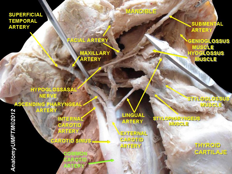
The internal carotid artery originates at the bifurcation of the common carotid artery, most commonly at the level of the superior border of the thyroid cartilage corresponding to the C3–C4 vertebral level.
This bifurcation represents one of the most important vascular junctions in the body, serving as the transition point between extracranial and intracranial circulatory systems. At its origin, the ICA lies posterolateral to the external carotid artery and is distinguished by its larger caliber, absence of cervical branches, and direct cranial destination.
The proximal ICA often exhibits a slight dilation known as the carotid sinus, which contains specialized baroreceptors responsible for monitoring systemic arterial pressure.
Adjacent to the bifurcation lies the carotid body, a highly vascular chemoreceptor organ that detects changes in blood oxygen, carbon dioxide, and pH. Together, these structures form a crucial neurovascular regulatory complex involved in cardiovascular homeostasis.
Embryologically, the ICA develops primarily from the third aortic arch and the cranial segment of the dorsal aorta. This developmental origin establishes the ICA as one of the earliest arterial pathways supplying the developing forebrain.
Exam Question
Evaluate the anatomical and physiological significance of the carotid bifurcation as the origin of the internal carotid artery, including the roles of the carotid sinus and carotid body in cardiovascular homeostasis.
Cervical Part
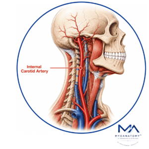
The cervical segment extends from the carotid bifurcation to the external opening of the carotid canal. Within the carotid sheath, the ICA ascends vertically through the neck accompanied by the internal jugular vein and vagus nerve.
Unlike the external carotid artery, it gives no cervical branches, emphasizing its exclusive role in supplying intracranial structures.
Throughout this segment, the artery remains closely related to the pharyngeal wall, superior cervical sympathetic ganglion, carotid sinus, and deep cervical fascial planes.
The absence of branches reduces turbulence and allows efficient transmission of blood toward the cranial cavity.
Because this segment is a frequent site of atherosclerotic plaque formation, it possesses major clinical relevance in ischemic cerebrovascular disease.
Exam Question
Discuss the anatomical course and relationships of the cervical segment of the internal carotid artery, and explain why this region is particularly susceptible to atherosclerotic disease and ischemic stroke.
Petrosus Part
The petrous segment begins as the ICA enters the carotid canal of the petrous temporal bone. Within this osseous tunnel, the vessel follows a curved course consisting of vertical and horizontal portions.
The artery is accompanied by the carotid sympathetic plexus and separated from surrounding bone by a venous plexus.
This segment is anatomically related to the middle ear cavity, cochlea, auditory tube, and inner ear structures. Consequently, fractures of the temporal bone, skull-base tumors, and otologic procedures may place the vessel at risk.
The petrous segment represents the transition between extracranial and intracranial environments and serves as a critical landmark in skull-base anatomy.
Exam Question
Assess the importance of the petrous segment as the transition between extracranial and intracranial circulation, and explain how its relations to the temporal bone and middle ear influence clinical risk during skull-base pathology.
Cavernous Part
After emerging from the petrous temporal bone, the ICA enters the cavernous sinus, where it forms the characteristic carotid siphon.
This S-shaped curvature modifies hemodynamic forces before blood enters the cerebral circulation and is believed to reduce pulsatile energy transmitted to delicate intracranial vessels.
The cavernous segment is surrounded by venous channels and exhibits exceptionally important neural relationships. The abducens nerve (CN VI) lies immediately adjacent to the artery, whereas cranial nerves III, IV, V1, and V2 occupy the lateral wall of the cavernous sinus.
Because of these relationships, aneurysms, thrombosis, inflammatory disease, or tumors affecting this region frequently produce combined neurovascular syndromes characterized by ophthalmoplegia, facial sensory deficits, and autonomic dysfunction.
Exam Question
Analyze the neurovascular significance of the cavernous segment of the internal carotid artery and explain how pathology within the cavernous sinus can simultaneously affect cerebral blood flow and multiple cranial nerves.
Cerebral Part
The supraclinoid segment begins after the ICA pierces the dura mater and enters the subarachnoid space.
This represents the terminal intracranial portion of the artery and is closely related to the optic nerve, optic chiasm, hypothalamus, pituitary stalk, and anterior perforated substance.
Within this segment, the ICA gives rise to several critical branches, including the ophthalmic artery, posterior communicating artery, and anterior choroidal artery.
These branches supply the orbit, retina, deep cerebral nuclei, internal capsule, and portions of the ventricular system. The supraclinoid segment therefore represents a major distribution center for both cortical and deep cerebral circulation.
Exam Question
Evaluate the role of the supraclinoid segment as the principal intracranial distribution center of the internal carotid artery, emphasizing its branches, target structures, and contribution to forebrain vascularization.
Termination
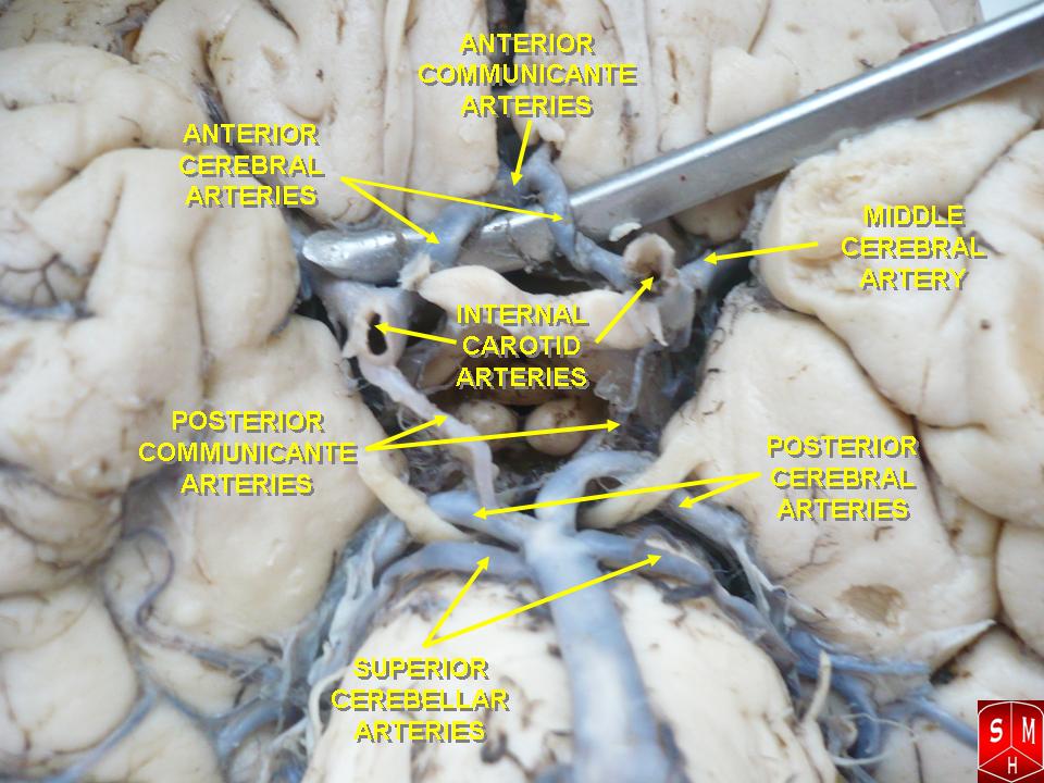
The internal carotid artery terminates near the anterior perforated substance, lateral to the optic chiasm and inferior to the frontal lobe. At this point, it divides into its two terminal branches: the anterior cerebral artery (ACA) and the middle cerebral artery (MCA).
These terminal vessels collectively supply the majority of the cerebral hemispheres. The ACA primarily vascularizes the medial surfaces of the frontal and parietal lobes, whereas the MCA supplies extensive lateral cortical territories involved in motor control, sensation, language, cognition, and higher cortical functions. Because these regions contain many of the most functionally important areas of the brain, interruption of ICA flow may result in profound neurological deficits.
Beyond simple distribution of blood, the terminal ICA forms a central component of the Circle of Willis, linking anterior and posterior cerebral circulations through communicating arteries. This arrangement provides an important collateral mechanism that may preserve cerebral perfusion during arterial stenosis, occlusion, or vascular injury.
Exam Question
Discuss the functional significance of the terminal bifurcation of the internal carotid artery into the anterior and middle cerebral arteries, and explain how this arrangement contributes to cerebral perfusion and collateral circulation through the Circle of Willis.
Anterior Relations
Anteriorly, the ICA is related to the posterior belly of the digastric muscle, stylohyoid muscle, and superior portions of the parotid region during its cervical ascent.
Intracranially, it becomes closely associated with the optic nerve and optic chiasm.
These relationships explain why vascular lesions of the ICA may produce visual disturbances and compressive optic neuropathies.
Exam Question
Explain how the anterior anatomical relationships of the internal carotid artery influence surgical access and discuss the potential visual consequences of pathology involving its intracranial anterior relations.
Posterior Relations
Posteriorly, the artery relates to the cervical sympathetic trunk, superior cervical ganglion, prevertebral fascia, and longus capitis muscle.
These structures contribute to autonomic regulation and provide important surgical landmarks during carotid exposure.
Exam Question
Evaluate the importance of the posterior relations of the internal carotid artery, particularly the cervical sympathetic trunk and superior cervical ganglion, in neurovascular regulation and carotid surgery.
Medial Relations
Medially, the ICA is related to the pharyngeal wall, laryngeal structures, and upper cervical viscera.
Within the cranial cavity, it lies adjacent to the pituitary gland, cavernous sinus, sphenoid sinus, and parasellar compartment.
These relationships are critically important in endoscopic skull-base surgery and pituitary procedures.
Exam Question
Analyze the clinical importance of the medial relations of the internal carotid artery within both the neck and cranial cavity, with particular reference to skull-base and pituitary procedures.
Lateral Relations
Laterally, the ICA is accompanied by the internal jugular vein and vagus nerve within the carotid sheath. In the cavernous sinus region, it becomes intimately associated with cranial nerves III, IV, V1, V2, and VI.
This dense concentration of neural structures makes the cavernous ICA one of the most clinically significant neurovascular crossroads in the human body.
Exam Question
Discuss why the lateral relations of the internal carotid artery constitute one of the most important neurovascular crossroads in the human body, emphasizing its association with the internal jugular vein, vagus nerve, and cavernous sinus cranial nerves.
SUMMARY TABLE