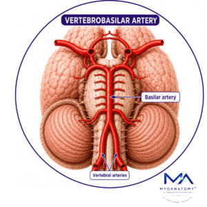
Vertebrobasilar Artery
MYO CORE
Clinical Relevance
Because numerous vital neurological centers are concentrated within the vertebrobasilar territory, even localized vascular compromise may produce disproportionate neurological dysfunction, making the posterior circulation one of the most clinically critical regions of the central nervous system.
OVERVIEW
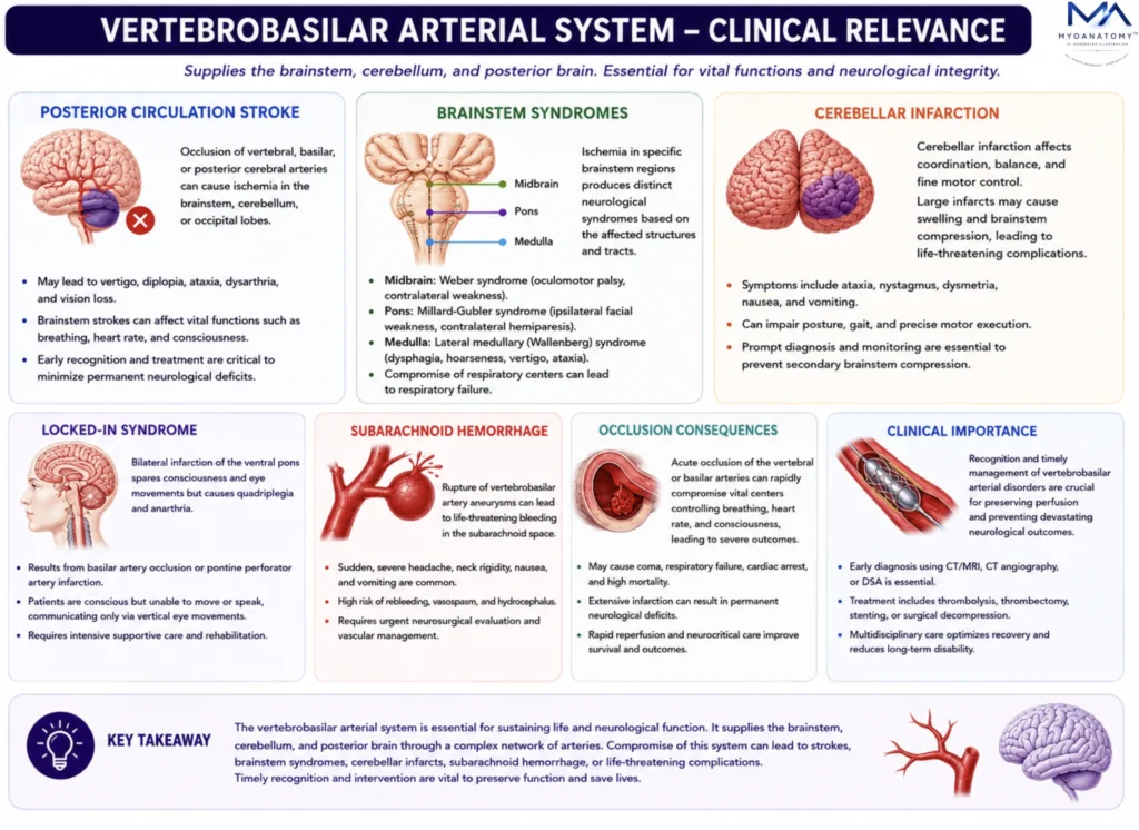
Consequently, vertebrobasilar vascular compromise may simultaneously disrupt consciousness, autonomic regulation, motor control, sensory transmission, balance, swallowing, ocular movements, hearing, and vision. Because these critical structures are densely concentrated within relatively small anatomical regions and often possess limited collateral circulation, even focal ischemia may result in severe neurological dysfunction.
Clinical manifestations range from vertigo, ataxia, diplopia, dysarthria, dysphagia, and hearing loss to devastating complications including posterior circulation stroke, brainstem infarction, locked-in syndrome, coma, respiratory failure, and sudden death.
As a result, vertebrobasilar pathology represents one of the most important causes of life-threatening neurological injury in clinical neurology, stroke medicine, and neurosurgery.
Exam Question
Assess the relationship between vertebrobasilar vascular anatomy and neurological function, and explain why focal ischemia within the posterior circulation may produce disproportionately severe clinical consequences ranging from isolated cranial nerve dysfunction to brainstem failure, locked-in syndrome, coma, and death.
CLINICAL RELEVANCE
Posterior Circulation Stroke
The vertebrobasilar arterial system supplies the brainstem, cerebellum, thalamus, occipital lobes, and upper cervical spinal cord. Consequently, occlusion of the vertebral, basilar, or posterior cerebral arteries may produce posterior circulation stroke, a major form of ischemic cerebrovascular disease.
Unlike anterior circulation infarction, vertebrobasilar ischemia frequently affects structures responsible for consciousness, ocular movements, balance, coordination, swallowing, autonomic regulation, and vision.
Clinical manifestations commonly include vertigo, diplopia, dysarthria, dysphagia, nystagmus, ataxia, visual field defects, cranial nerve deficits, sensory disturbances, limb weakness, and impaired consciousness.
Because numerous critical neural pathways converge within a compact anatomical region, even relatively small infarctions may simultaneously disrupt multiple neurological systems.
Posterior circulation stroke therefore constitutes a neurological emergency due to its potential to compromise vital brainstem functions essential for survival.
Exam Question
Analyze how the anatomical territories supplied by the vertebrobasilar circulation explain the diverse neurological manifestations of posterior circulation stroke, including disturbances of consciousness, ocular motility, balance, coordination, swallowing, autonomic regulation, and visual processing.
Brainstem Syndromes
The brainstem contains a dense concentration of cranial nerve nuclei, reticular activating pathways, autonomic control centers, ascending sensory tracts, and descending motor pathways.
As a result, ischemia involving vertebral, basilar, or perforating arteries may produce highly characteristic neurological syndromes that reflect the precise vascular territory affected.
Major vertebrobasilar syndromes include lateral medullary (Wallenberg), medial medullary, pontine, and midbrain infarction syndromes.
Patients may present with dysphagia, dysarthria, hoarseness, facial sensory loss, contralateral body sensory deficits, gaze abnormalities, limb weakness, ataxia, respiratory dysfunction, and disturbances of consciousness.
Because many brainstem perforators function as end arteries with limited collateral circulation, even small focal infarctions may generate severe and predictable neurological deficits.
Exam Question
Evaluate how the compact arrangement of cranial nerve nuclei, ascending sensory pathways, descending motor tracts, reticular activating networks, and autonomic centers within the brainstem contributes to the characteristic clinical syndromes produced by vertebrobasilar ischemia.
Cerebellar Infarction
Occlusion of the PICA, AICA, or SCA may produce cerebellar infarction involving the cerebellar hemispheres, vermis, deep cerebellar nuclei, and cerebellar peduncles. These structures are essential for motor coordination, balance, postural control, movement refinement, and motor learning.
Clinical manifestations include gait instability, truncal or limb ataxia, dysmetria, intention tremor, dysarthria, nystagmus, vertigo, and impaired equilibrium. Because cerebellar infarction often presents with dizziness and balance disturbance rather than obvious paralysis, diagnosis may be delayed.
Large cerebellar infarctions are particularly dangerous because edema within the posterior cranial fossa may compress the fourth ventricle and brainstem, causing obstructive hydrocephalus, secondary brainstem compression, herniation, and potentially fatal neurological deterioration.
Exam Question
Assess how interruption of blood flow through the PICA, AICA, or SCA affects cerebellar hemispheres, vermis, deep cerebellar nuclei, and cerebellar peduncles, and explain the resulting disturbances of coordination, balance, posture, motor learning, and movement refinement.
Locked In Syndrome
Locked-in syndrome is one of the most devastating consequences of vertebrobasilar disease and most commonly results from basilar artery occlusion causing bilateral infarction of the ventral pons. The lesion destroys corticospinal and corticobulbar pathways that convey voluntary motor commands from the cerebral cortex.
Despite profound motor paralysis, the reticular activating system and cerebral cortex remain largely intact.
Consequently, patients preserve consciousness, awareness, cognition, and sensory perception while losing nearly all voluntary motor function.
Affected individuals typically develop quadriplegia, anarthria, facial paralysis, and inability to swallow or communicate verbally.
Vertical eye movements and blinking are often preserved because their controlling pathways remain unaffected, providing the patient’s primary means of communication.
Exam Question
Analyze the neuroanatomical basis of locked-in syndrome and explain how basilar artery occlusion can selectively disrupt corticospinal and corticobulbar pathways while preserving consciousness, cognition, and sensory awareness.
Subarachnoid Hemorrhage
Subarachnoid hemorrhage (SAH) is a life-threatening neurological emergency caused by bleeding into the subarachnoid space, most commonly following rupture of an intracranial aneurysm. Vertebrobasilar aneurysms are particularly dangerous because of their close relationship to the brainstem and adjacent neurovascular structures.
Extravasated blood rapidly increases intracranial pressure, impairs cerebral perfusion, and triggers secondary neuronal injury.
Patients typically present with sudden severe thunderclap headache, nausea, vomiting, photophobia, neck stiffness, altered consciousness, and focal neurological deficits. Major complications include cerebral vasospasm, delayed cerebral ischemia, hydrocephalus, rebleeding, seizures, brain herniation, and brainstem compression.
Consequently, rupture of a posterior circulation aneurysm may rapidly progress to respiratory failure, coma, or death.
Exam Question
Analyze how rupture of a vertebrobasilar aneurysm produces both primary neurological injury and secondary complications, and explain why subarachnoid hemorrhage may result in cerebral ischemia, hydrocephalus, and life-threatening brainstem dysfunction.
Occlusion Consequence
Occlusion of the vertebral, basilar, or posterior cerebral arteries may compromise perfusion of the brainstem, cerebellum, thalamus, occipital lobes, and upper cervical spinal cord.
Because these territories contain respiratory and cardiovascular centers, reticular activating pathways, cranial nerve nuclei, major sensorimotor tracts, and cerebellar networks, even focal ischemia may produce profound neurological dysfunction.
Clinical manifestations commonly include vertigo, diplopia, dysarthria, dysphagia, ataxia, nystagmus, visual disturbances, sensory deficits, limb weakness, and impaired consciousness.
Severe vertebrobasilar occlusion may cause extensive brainstem infarction, locked-in syndrome, coma, respiratory failure, or death. The severity of these deficits reflects the dense concentration of vital neural structures and the limited collateral circulation within many posterior circulation territories.
Exam Question
Evaluate how vertebrobasilar arterial occlusion disrupts brainstem, cerebellar, and posterior cerebral function, and explain why relatively small ischemic lesions may produce disproportionately severe neurological deficits.
SUMMARY TABLE