Vertebrobasilar Artery
MYO CORE
Branches & Distribution
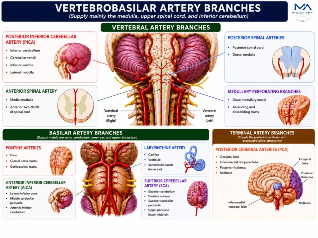
The vertebrobasilar system distributes blood through the vertebral and basilar arteries and their major branches, establishing the principal arterial network of the posterior circulation. Together, these vessels provide coordinated perfusion to the brainstem, cerebellum, posterior cerebrum, and upper spinal cord.
OVERVIEW
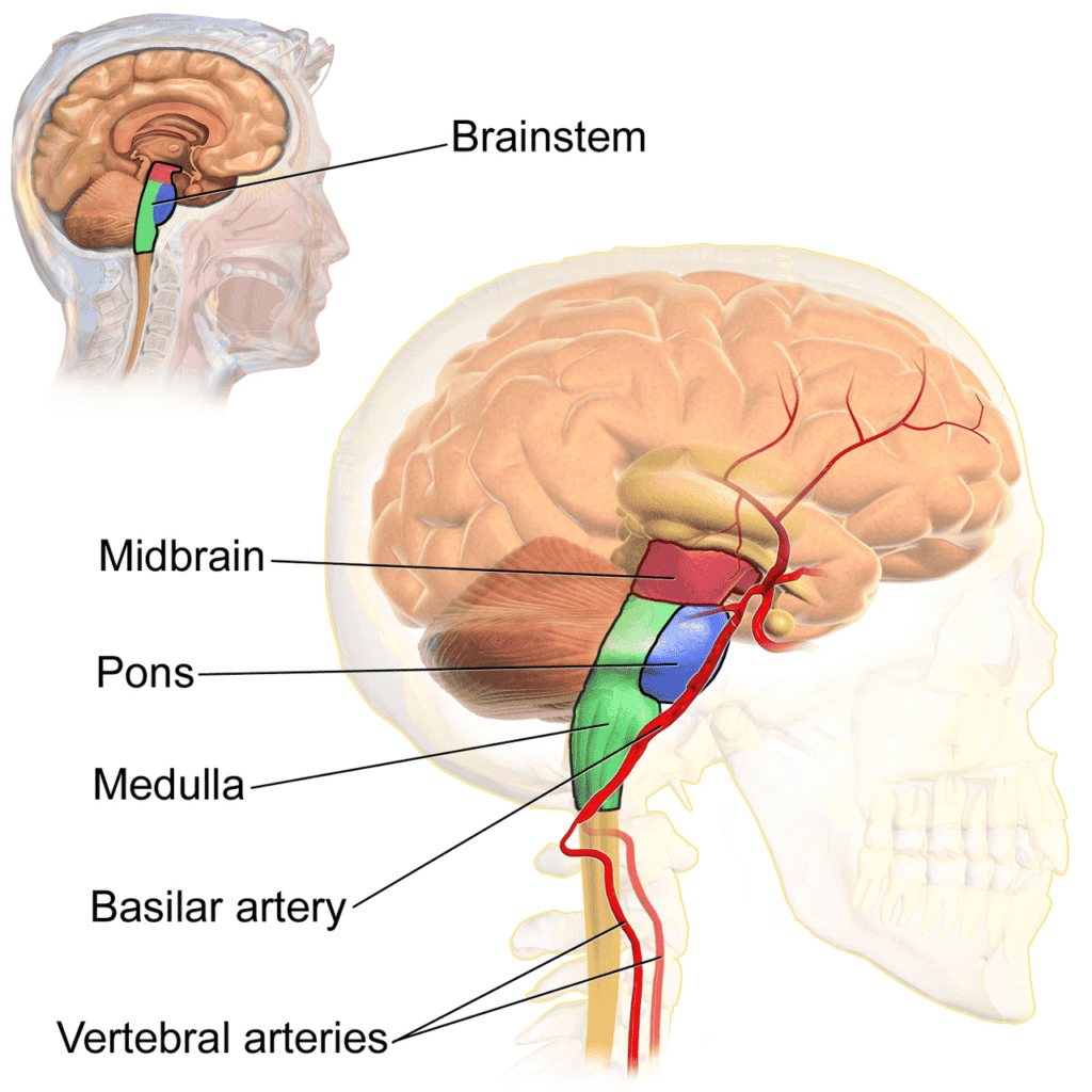
The vertebral arteries form the foundational vessels of the posterior circulation and supply the medulla oblongata, upper cervical spinal cord, inferior cerebellum, and numerous autonomic and sensorimotor centers essential for life.
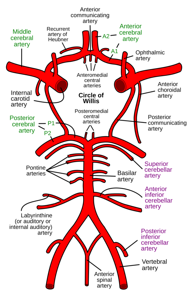
Their branches include the Posterior Inferior Cerebellar Artery (PICA), Anterior Spinal Artery, Posterior Spinal Arteries, and multiple medullary perforating branches.
Together, these vessels vascularize structures responsible for respiration, cardiovascular regulation, swallowing, balance, coordination, and cranial nerve function.
Because these neurological structures are densely concentrated within the medulla, even small vertebral artery lesions may produce profound neurological deficits.
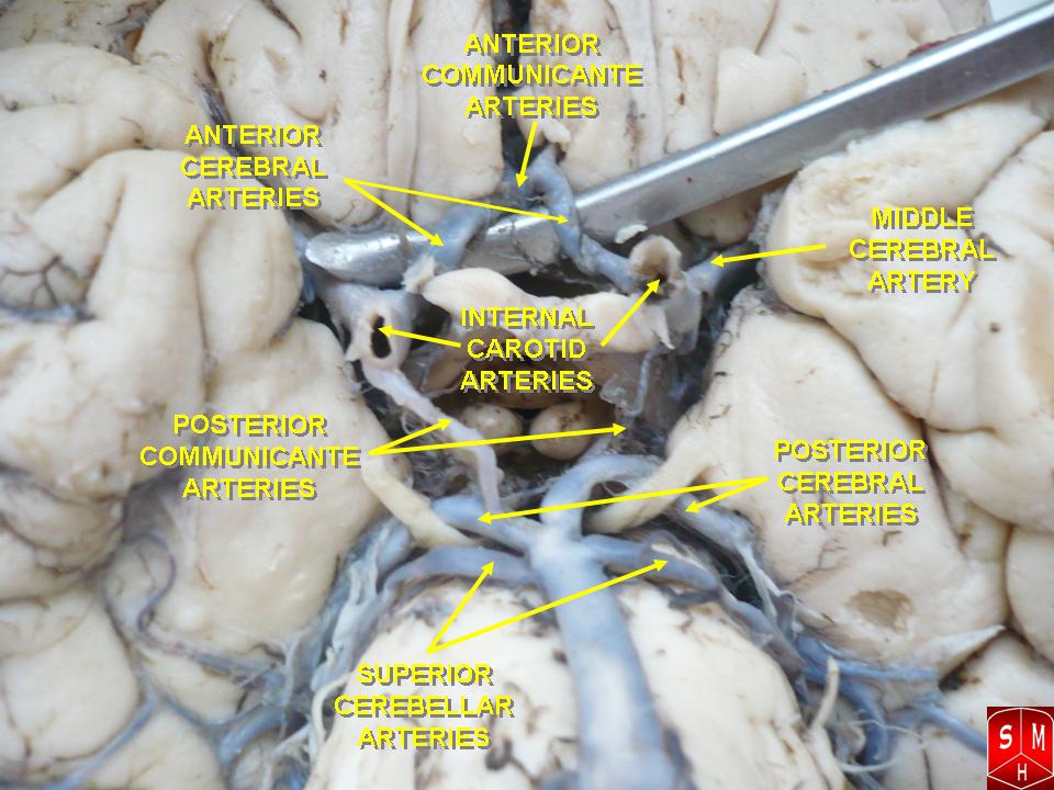
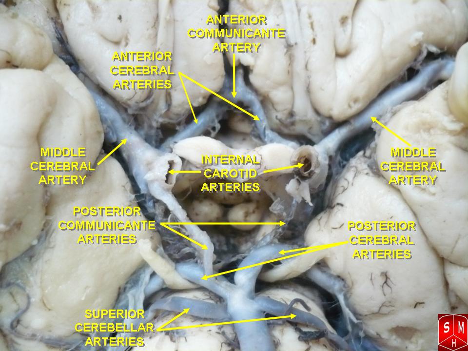
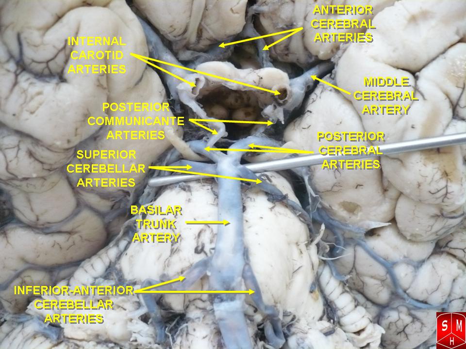
Vertebral Artery Branches
Posterior Inferior Cerebellar Artery
The posterior inferior cerebellar artery is typically the largest branch of the intracranial vertebral artery and represents the principal arterial supply to the inferior cerebellum and dorsolateral medulla.
After arising near the vertebrobasilar junction, it follows a tortuous course around the lateral medulla before entering the cerebellomedullary fissure, where it divides into multiple hemispheric and vermian branches.
Its territory includes the inferior cerebellar hemisphere, cerebellar tonsil, inferior vermis, and extensive portions of the lateral medulla. Through numerous perforating branches, PICA supplies the vestibular nuclei, nucleus ambiguus, spinal trigeminal nucleus, inferior cerebellar peduncle, spinothalamic tract, reticular formation, and descending sympathetic pathways.
Functionally, this vascular territory integrates equilibrium, coordination, swallowing, phonation, autonomic regulation, and sensory processing.
Consequently, PICA represents one of the most clinically significant arteries of the posterior circulation because ischemia may simultaneously disrupt multiple neurological systems.
Exam Question
Analyze how the anatomical distribution of the PICA enables simultaneous vascular supply to the cerebellum, vestibular system, and lateral medulla, and explain the functional significance of this arrangement.
Anterior Spinal Artery
The anterior spinal artery originates from paired vertebral contributions near the lower medulla and descends within the anterior median fissure of the spinal cord.
Although commonly regarded as a spinal vessel, its proximal branches form a major component of vertebrobasilar circulation through their supply to the medial medulla.
Its territory includes the pyramids, medial lemniscus, hypoglossal nucleus, medial reticular formation, and anterior two-thirds of the spinal cord.
These structures contain corticospinal fibers responsible for voluntary movement, ascending sensory pathways transmitting proprioceptive information, and motor nuclei controlling tongue movements.
The anterior spinal artery therefore serves as the principal vascular conduit linking brainstem motor pathways with the spinal cord while maintaining perfusion of critical medullary structures involved in motor execution and sensory integration.
Exam Question
Evaluate the importance of the anterior spinal artery as a vascular link between the medulla oblongata and spinal cord, with particular reference to the neural pathways contained within its territory.
Posterior Spinal Artery
The posterior spinal arteries arise either directly from the vertebral arteries or indirectly through PICA.
They descend along the posterolateral surface of the spinal cord and provide blood supply to the posterior spinal cord and dorsal medulla.
Their branches vascularize the gracile and cuneate nuclei, dorsal columns, posterior horns, and adjacent sensory relay centers responsible for fine touch, vibration, and conscious proprioception.
Although smaller than the anterior spinal artery, the posterior spinal arteries maintain the integrity of sensory systems essential for posture, balance, coordinated movement, and spatial orientation.
Exam Question
Explain how the vascular territory of the posterior spinal arteries supports dorsal column function and contributes to sensory integration within the central nervous system.
Medullary Perforating Artery
Numerous small perforating arteries arise directly from the vertebral arteries and penetrate the medulla oblongata.
Despite their small size, these vessels supply some of the most vital neural structures within the entire nervous system.
Their territories include respiratory centers, cardiovascular regulatory nuclei, reticular activating pathways, lower cranial nerve nuclei, corticospinal tracts, spinothalamic pathways, and medial lemniscal systems.
Because collateral circulation within these deep medullary regions is limited, even focal occlusion of a single perforating vessel may produce severe neurological dysfunction.
These branches therefore play a disproportionately important role in maintaining consciousness, autonomic homeostasis, sensorimotor integration, and life-sustaining brainstem functions.
Exam Question
Assess the functional importance of vertebral medullary perforating arteries in maintaining autonomic regulation, sensorimotor transmission, and brainstem homeostasis.
Basilar Artery Branches
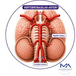
The basilar artery is formed by the union of the right and left vertebral arteries at the pontomedullary junction and serves as the central arterial trunk of the posterior circulation. Its major branches include the pontine arteries, anterior inferior cerebellar artery (AICA), labyrinthine artery, and superior cerebellar artery (SCA), before terminating as the right and left posterior cerebral arteries (PCA). Together, these vessels supply the pons, cerebellum, inner ear, upper brainstem, and numerous cranial nerve nuclei.
Collectively, they support motor control, sensory transmission, ocular movements, auditory processing, vestibular integration, cerebellar coordination, and higher visual functions within the posterior cerebrum.
Pontine Arteries
The pontine arteries are the most numerous branches of the basilar artery and may be classified into paramedian, short circumferential, and long circumferential groups.
These vessels penetrate the substance of the pons and supply corticospinal tracts, corticobulbar pathways, medial lemniscus fibers, pontine nuclei, reticular formation, and multiple cranial nerve nuclei.
Functionally, they maintain communication between the cerebral cortex, cerebellum, spinal cord, and brainstem. Their territories contain pathways essential for voluntary movement, facial expression, ocular coordination, sensory processing, and consciousness.
Because of the dense arrangement of neurological structures within the pons, pontine arterial lesions often produce extensive neurological impairment.
Exam Question
Analyze how the pontine arteries sustain communication between the cerebral cortex, cerebellum, spinal cord, and cranial nerve nuclei through their vascular supply of the pons.
Anterior Inferior Cerebellar Artery
The anterior inferior cerebellar artery typically arises from the lower basilar artery and courses laterally around the pontomedullary region toward the cerebellopontine angle.
Its vascular territory includes the lateral inferior pons, middle cerebellar peduncle, flocculus, and anterior inferior cerebellar hemisphere.
Through its perforating branches it supplies structures involved in balance, coordination, vestibular processing, facial motor control, and auditory function.
The middle cerebellar peduncle, supplied largely by AICA, constitutes a major afferent pathway connecting the cerebral cortex to the cerebellum.
Consequently, AICA serves as an important vascular bridge between pontine and cerebellar functional systems.
Exam Question
Trace the vascular territory of the AICA and explain how its anatomical distribution integrates cerebellar, vestibular, auditory, and facial motor functions.
Labyrinth Artery
The labyrinthine artery usually arises from AICA, although direct origin from the basilar artery may occur.
It accompanies the facial and vestibulocochlear nerves through the internal acoustic meatus and constitutes the exclusive arterial supply of the membranous labyrinth.
Its branches supply the cochlea, vestibule, semicircular canals, utricle, and saccule. These structures contain the specialized sensory receptors responsible for hearing and equilibrium.
Because the labyrinthine artery behaves as a functional end artery with minimal collateral circulation, interruption of blood flow may rapidly injure highly metabolically active sensory neuroepithelium.
Consequently, it plays a critical role in maintaining auditory and vestibular function.
Exam Question
Evaluate the significance of the labyrinthine artery as the principal arterial supply of the membranous labyrinth and discuss the functional implications of its limited collateral circulation.
Superior Cerebellar Artery
The superior cerebellar artery arises immediately inferior to the terminal bifurcation of the basilar artery and courses around the cerebral peduncle beneath the oculomotor nerve.
Its territory includes the superior cerebellar hemisphere, superior vermis, dentate nucleus, superior cerebellar peduncle, upper pons, and lower midbrain.
These structures constitute the principal output pathways of the cerebellum and are responsible for motor planning, movement refinement, postural regulation, and coordination of voluntary activity.
Through its brainstem perforators, the SCA additionally contributes to vascularization of neural networks involved in ocular movements and sensorimotor integration.
The superior cerebellar artery therefore represents the dominant vascular supply to the cerebellar efferent system.
Exam Question
Analyze how the superior cerebellar artery supports cerebellar output systems involved in motor planning, movement refinement, and postural coordination.
Terminal Artery Branches
The posterior cerebral arteries (PCA) represent the terminal branches of the basilar artery and form the principal arterial supply of the posterior cerebrum. Their major branches include cortical branches to the occipital and temporal lobes, thalamic perforating branches, posterior choroidal arteries, and midbrain perforators.
Together, these vessels supply the occipital cortex, inferomedial temporal lobe, posterior thalamus, midbrain, splenium of the corpus callosum, and visual association areas.
Collectively, they support visual perception, visual interpretation, memory processing, sensory integration, thalamocortical communication, and higher-order cognitive functions associated with the posterior cerebral hemispheres. Because the PCA supplies both cortical and deep diencephalic structures, vascular compromise may simultaneously affect vision, memory, consciousness, and complex neurocognitive processing.
SUMMARY TABLE