Epicranial Muscle
MYO CORE
Auricularis Muscle
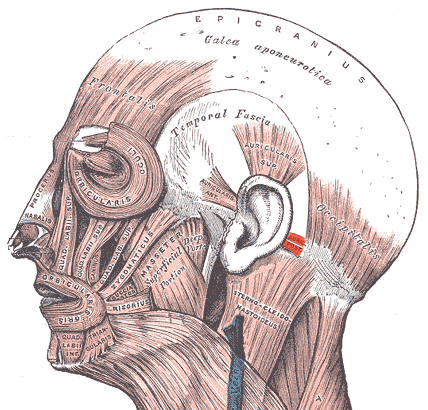
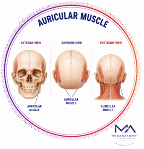
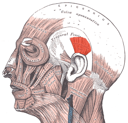
The auricular muscles are a small group of muscles of facial expression associated with movement of the external ear (auricle). They consist of three extrinsic muscles:
Auricularis anterior
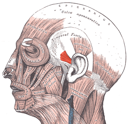
Auricularis superior
Auricularis posterior
OVERVIEW
These muscles connect the auricle to the scalp and temporal region, allowing limited movements of the ear. In many mammals these muscles are highly developed and permit substantial ear movement for sound localization.
In humans, however, they are rudimentary and functionally weak, reflecting the reduced importance of auricular mobility in human auditory function.
Anatomically, the auricular muscles lie within the superficial fascia of the scalp and temporal region, closely associated with the epicranial aponeurosis and surrounding facial expression musculature. They act primarily to stabilize and slightly reposition the auricle, rather than producing large movements.
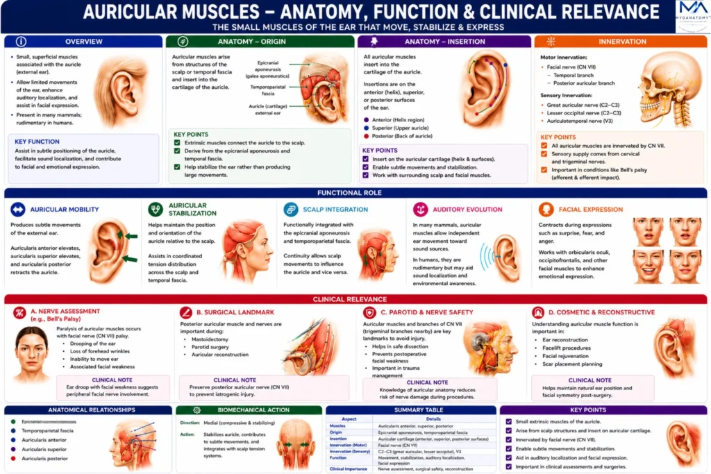
The auricular muscles – auricularis anterior, superior, and posterior- are small extrinsic muscles connecting the auricle to the scalp. Although functionally reduced in humans, they permit minor movements of the external ear and contribute to stabilization of auricular position.
They derive from the second pharyngeal arch and are innervated by branches of the facial nerve, linking them anatomically to the broader system of facial expression musculature.
Their clinical importance lies primarily in their relationship to facial nerve branches and surgical anatomy of the auricular and mastoid regions.
Although their functional role in humans is limited, they remain anatomically important because of their relationships with facial nerve branches, scalp fascia, and auricular structures.

ANATOMY
Origin
Each auricular muscle arises from structures of the scalp or temporal fascia.
Auricularis anterior -temporal fascia and epicranial aponeurosis
Auricularis superior- epicranial aponeurosis (galea aponeurotica)
Auricularis posterior – mastoid process of the temporal bone
Exam Question
Compare the origins of the auricularis anterior, auricularis superior, and auricularis posterior muscles, and explain how their distinct anatomical attachments influence the direction of auricular movement.
Insertion
The muscles insert into the cartilage of the auricle.
Auricularis anterior – onto anterior surface of the auricle (helix region)
Auricularis superior – onto superior portion of the auricle
Auricularis posterior – onto posterior surface of the auricle
Exam Question
Describe the insertion sites of the three auricular muscles and analyze how their insertions into different regions of the auricular cartilage determine their specific biomechanical actions on the external ear.
Innervation
All auricular muscles receive motor innervation from the facial nerve (cranial nerve VII).
Auricularis anterior – temporal branches of the facial nerve
Auricularis superior – temporal branches of the facial nerve
Auricularis posterior – posterior auricular branch of the facial nerve
The posterior auricular nerve arises from the facial nerve shortly after it exits the stylomastoid foramen.
Exam Question
Explain the motor innervation of the auricular muscles, detailing the contributions of the temporal and posterior auricular branches of the facial nerve (CN VII), and discuss the anatomical significance of the posterior auricular nerve following its emergence from the stylomastoid foramen.
FUNCTIONAL ROLE
Auricular Mobility
The auricular muscles produce subtle movements of the external ear (auricle). The auricularis anterior draws the auricle anteriorly, the auricularis superior elevates it superiorly, and the auricularis posterior retracts it posteriorly.
Although these movements are minimal in most humans, they represent the residual motor capacity of the external ear
Exam Question
Evaluate the individual actions of the auricularis anterior, superior, and posterior muscles and discuss why these movements remain rudimentary in most humans despite preserved motor innervation
Auricular Stabilization
The auricular muscles help maintain the position and orientation of the auricle relative to the scalp.
During movements generated by the occipitofrontalis muscle, they provide dynamic stabilization, preventing excessive displacement of the external ear and contributing to coordinated scalp–auricle mechanics.
Exam Question
Discuss the role of the auricular muscles in maintaining auricular position during scalp movement and explain the mechanisms by which they contribute to dynamic stabilization of the external ear.
Scalp Integration
Functionally, the auricular muscles are integrated with the epicranial musculoaponeurotic system through their attachments to the epicranial aponeurosis (galea aponeurotica) and temporal fascia.
This anatomical continuity allows scalp movements to influence auricular position and facilitates coordinated tension distribution across the lateral cranial soft tissues.
Exam Question
Analyze the anatomical and functional relationship between the auricular muscles, the epicranial aponeurosis (galea aponeurotica), and the temporal fascia, emphasizing their integration within the epicranial musculoaponeurotic system
Auditory Evolution
In numerous mammals, the auricular muscles permit independent ear movements toward sound sources, significantly enhancing auditory localization and environmental awareness.
In humans, these muscles have undergone marked functional regression, with sound localization relying predominantly on head movement and binaural auditory processing rather than active auricular motion.
Exam Question
Critically evaluate the evolutionary regression of the human auricular muscles by comparing their function in humans with that in other mammals, particularly regarding sound localization and environmental awareness.
CLINICAL RELEVANCE
Nerve Assessment
The auricular muscles may be affected by lesions of the facial nerve (CN VII), particularly involving the posterior auricular branch.
Although loss of auricular movement rarely produces functional impairment, weakness or paralysis of these muscles can serve as a subtle clinical indicator of facial nerve dysfunction and aid neurological assessment.
Exam Question
Explain how dysfunction of the auricular muscles may assist in the neurological assessment of facial nerve lesions and discuss why weakness of these muscles may provide valuable localizing information despite limited functional impairment.
Facial Paralysis
In peripheral facial nerve palsy, denervation of the auricular muscles may accompany weakness of other muscles of facial expression. Evaluation of auricular muscle function can provide additional information regarding the extent and anatomical level of facial nerve involvement.
Exam Question
Discuss the clinical significance of auricular muscle involvement in peripheral facial nerve palsy and explain how assessment of auricular muscle function may help determine the anatomical extent of facial nerve injury
Surgical Landmark
The posterior auricular muscle and posterior auricular nerve constitute important anatomical landmarks during mastoid, parotid, and facial nerve surgeries.
Their identification assists surgeons in locating and preserving adjacent branches of the facial nerve, thereby reducing the risk of iatrogenic nerve injury.
Exam Question
Describe the surgical importance of the posterior auricular muscle and posterior auricular nerve as anatomical landmarks during mastoid, parotid, and facial nerve procedures, emphasizing their role in reducing iatrogenic nerve injury.
Diagnostic Marker
Despite their limited functional role, the auricular muscles possess clinical value as anatomical and neurophysiological markers that can assist in evaluating facial nerve continuity, surgical orientation, and postoperative nerve recovery.
Exam Question
Evaluate the value of the auricular muscles as anatomical and neurophysiological markers in modern clinical practice, including their applications in facial nerve continuity assessment, surgical orientation, and postoperative recovery monitoring.
SUMMARY TABLE