The temporoparietalis muscle is a thin, superficial muscle of facial expression located within the lateral scalp, extending over the temporal region between the auricle and the epicranial aponeurosis. It represents a lateral continuation of the epicranial (scalp) musculature and is considered a vestigial remnant of the more highly developed auricular musculature found in many mammals.

Temporoparietalis Muscle

CORE

Overview

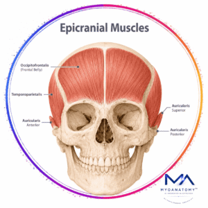
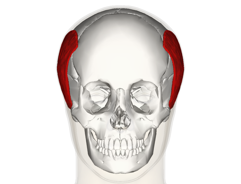
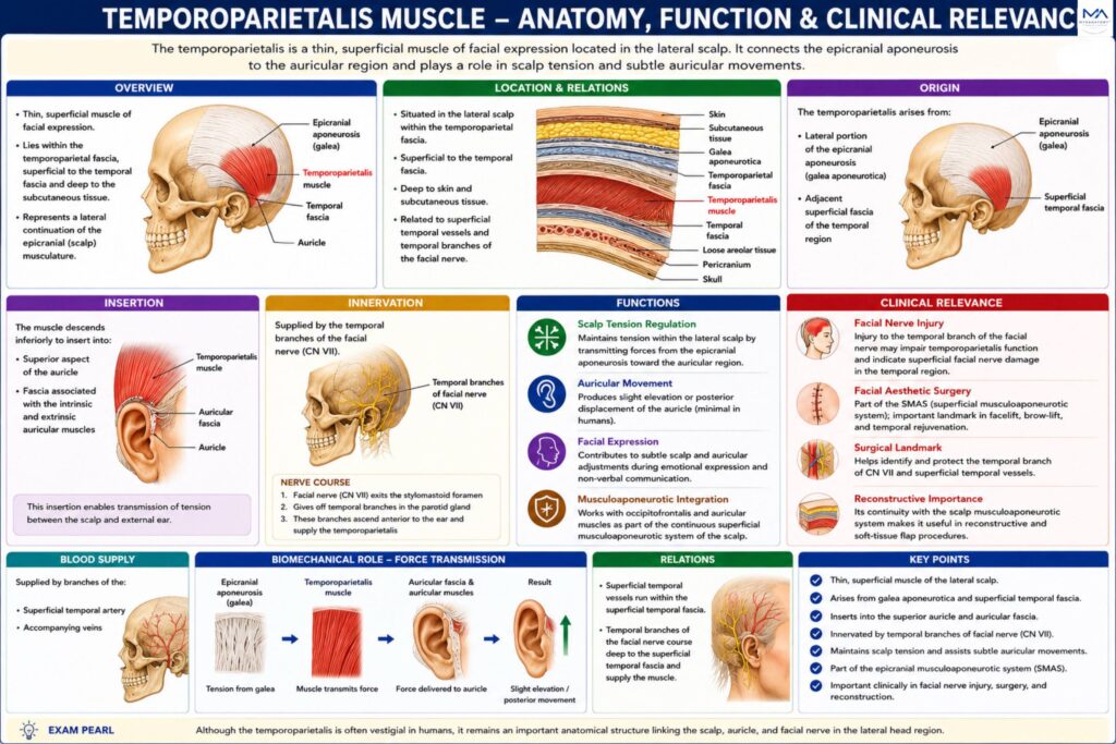
Situated within the temporoparietal fascia, the muscle lies superficial to the temporal fascia and deep to the skin and subcutaneous tissues of the scalp. Although relatively small and often poorly developed in humans, it serves as an important anatomical landmark because of its close relationship to the superficial temporal vessels and the temporal branches of the facial nerve.

Functionally, the temporoparietalis contributes to subtle movements of the scalp and external ear and participates in the coordinated musculoaponeurotic system of the lateral scalp.

ANATOMY

Origin

The temporoparietalis arises from:

lateral portion of the epicranial aponeurosis (galea aponeurotica)

adjacent superficial fascia of the temporal region

These attachments connect the muscle

Exam Question

Describe the origin of the temporoparietalis muscle. How do its attachments to the epicranial aponeurosis and temporal fascia anatomically link the scalp to the auricular region.

Insertion

The muscle descends inferiorly to insert into:

superior aspect of the auricle

fascia associated with the intrinsic and extrinsic auricular muscles

This insertion enables transmission of tension between the scalp and external ear.

Exam Question

Explain the insertion of the temporoparietalis muscle. How does its attachment to the auricular fascia facilitate transmission of tension between the scalp and external ear.

Innervation

The temporoparietalis is supplied by:

Temporal branches of the facial nerve (CN VII)

These branches provide motor innervation as part of the muscles of facial expression.

Exam Question

Identify the motor innervation of the temporoparietalis muscle. How would injury to the temporal branches of the facial nerve affect scalp mobility, auricular movement, and facial expression.

FUNCTIONAL ROLE

Scalp Tension Regulation

The temporoparietalis assists in maintaining tension within the lateral scalp by transmitting forces from the epicranial aponeurosis toward the auricular region

Exam Question

Explain the biomechanical role of the temporoparietalis in scalp tension regulation. How does its attachment to the epicranial aponeurosis contribute to force transmission and coordinated scalp movement.

Auricular Movement

Contraction may produce slight elevation or posterior displacement of the auricle.

Although this movement is minimal in most individuals, it represents a remnant of the highly mobile auricular musculature seen in other mammals

Exam Question

Analyze the function of the temporoparietalis in auricular movement. Why is this action considered a vestigial remnant of auricular musculature, and what is its evolutionary significance.

Epicranial Muscle Integration

The muscle acts synergistically with:

Occipitofrontalis

Auricular muscles

Temporoparietal fascia

Together they form a continuous superficial musculoaponeurotic layer across the scalp.

Exam Question

Discuss how the temporoparietalis integrates functionally with the occipitofrontalis, auricular muscles, and temporoparietal fascia. How do these structures form a continuous musculoaponeurotic system of the scalp.

Facial Expression

Through its connections with the scalp and auricle, the temporoparietalis contributes to subtle adjustments of facial and scalp positioning during emotional expression.


Exam Question

Evaluate the contribution of the temporoparietalis to facial expression. How do its anatomical connections influence subtle scalp and auricular movements during emotional communication.

CLINICAL RELEVANCE

Facial Nerve Injury


Damage to the temporal branch of the facial nerve may impair temporoparietalis function and serves as an indicator of superficial facial nerve injury in the temporal region.

Exam Question

Explain the functional consequences of injury to the temporal branch of the facial nerve on the temporoparietalis muscle. How can impairment of this muscle assist in the clinical assessment of superficial facial nerve lesions within the temporal region.

Facial Aesthetic Surgery

The temporoparietalis forms part of the superficial musculoaponeurotic system (SMAS) and serves as an important anatomical landmark during facelift, brow-lift, and temporal rejuvenation procedures. Preservation of the muscle helps maintain normal facial contour and soft-tissue mobility.

Exam Question

Discuss the anatomical relationship between the temporoparietalis muscle and the superficial musculoaponeurotic system (SMAS). Why is preservation of this structure important during facelift, brow-lift, and temporal rejuvenation procedures.

Surgical Landmark

The muscle provides a valuable surgical guide to deeper temporal structures, including the superficial temporal vessels and temporal branches of the facial nerve. Accurate identification reduces the risk of neurovascular injury during craniofacial and temporal surgery.

Exam Question

Why is the temporoparietalis muscle considered an important surgical landmark in the temporal region? Describe its relationship to the superficial temporal vessels and temporal branches of the facial nerve and explain its significance during craniofacial surgery.

Reconstructive Application

Due to its continuity with the scalp musculoaponeurotic system, the temporoparietalis may be incorporated into reconstructive and facial reanimation procedures to restore soft-tissue support, symmetry, and dynamic facial function

Exam Question

Evaluate the role of the temporoparietalis muscle in reconstructive and facial reanimation surgery. How does its continuity with the scalp musculoaponeurotic system contribute to restoration of facial symmetry and dynamic facial movement.

SUMMARY TABLE

Scroll to Top