The occipitofrontalis muscle, also known as the epicranius, is a broad paired muscle of facial expression that covers the superior aspect of the skull and the forehead. It is composed of two muscular bellies – the frontal belly (frontalis) and the occipital belly (occipitalis)- which are connected by a strong fibrous sheet called the epicranial aponeurosis (galea aponeurotica).

Occipitofrontalis Muscle

CORE

Overview

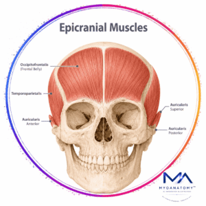
Together these structures form the musculoaponeurotic layer of the scalp, allowing coordinated movement of the scalp and forehead.

Occipitofrontalis lies within the superficial fascia of the scalp, deep to the skin and subcutaneous connective tissue but superficial to the loose areolar layer that separates the scalp from the skull.

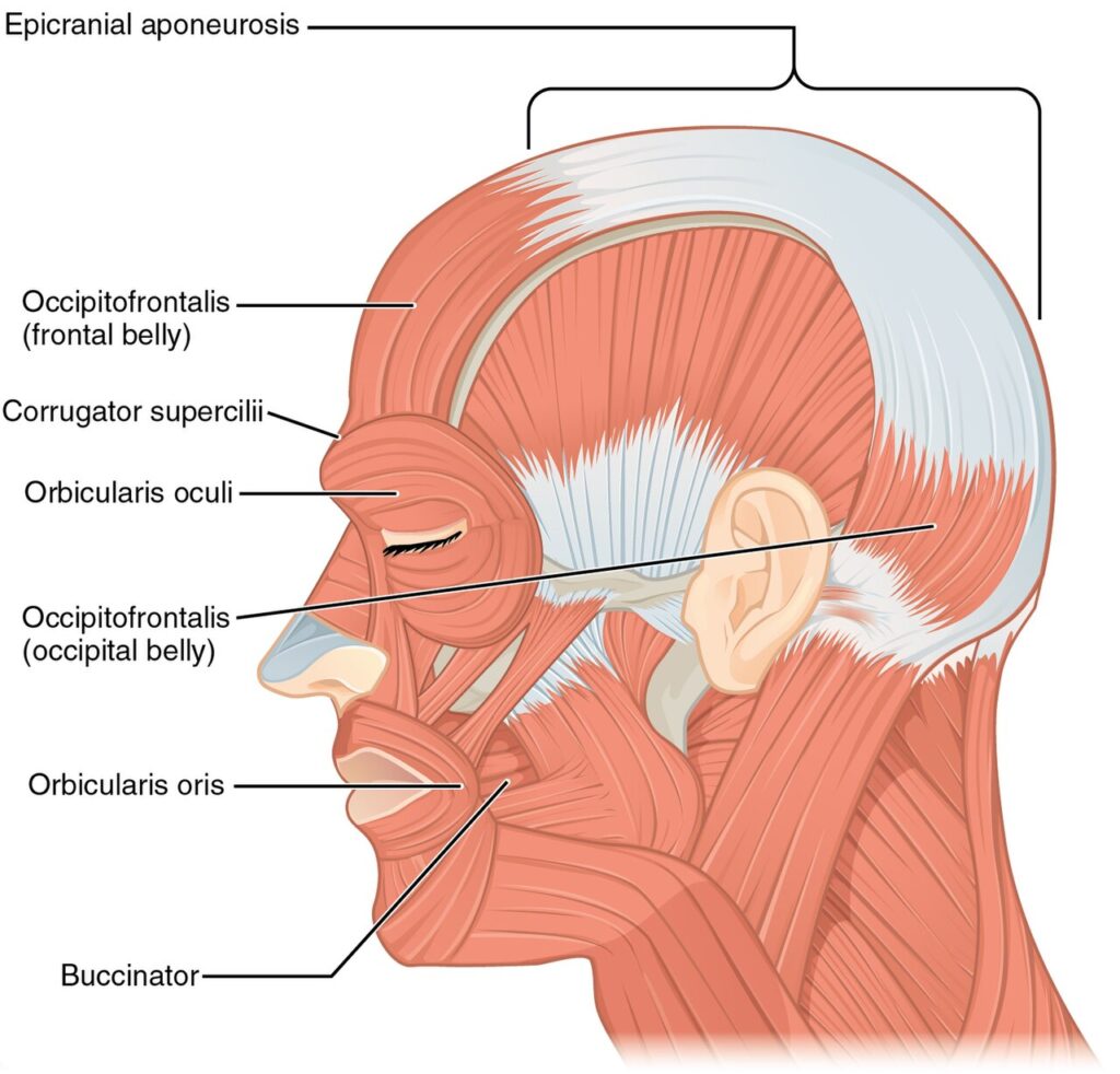
Unlike most skeletal muscles, the frontal portion of the occipitofrontalis inserts into the dermis of the forehead and eyebrows rather than bone, which enables direct movement of the skin. Through these attachments, the muscle produces visible changes in facial expression and contributes to the complex system of non-verbal emotional communication.

ANATOMY

Origin

Because the occipitofrontalis consists of two bellies connected by an aponeurosis, the muscle has two distinct points of origin.

Occipital Belly

originates from – lateral two-thirds of the superior nuchal line of the occipital bone; mastoid process of the temporal bone

Frontal belly 

originates from – gala aponeurotica

These attachments anchor the muscle to the posterior aspect of the skull.

Exam Question

Describe the anatomical attachments of the occipitofrontalis muscle. How do the origins of the occipital and frontal bellies enable coordinated movement of the scalp and forehead

Insertion

The occipital belly inserts into

epicranial aponeurosis (galea aponeurotica) From there

Frontal belly arises and inserts into:

skin and subcutaneous tissue of the forehead

skin of the eyebrows

root of the nose

This arrangement allows the muscle to act across the entire scalp-forehead complex.

Exam Question

Explain the insertion pattern of the occipitofrontalis muscle. Why is insertion of the frontal belly into the skin of the forehead and eyebrows functionally important for facial expression

Innervation

Both bellies are innervated by branches of the facial nerve (cranial nerve VII):

Frontal belly: temporal branch of the facial nerve

Occipital belly: posterior auricular branch of the facial nerve

These nerves provide motor control to the muscle as part of the mimetic musculature responsible for facial expression.

Exam Question

Identify the motor innervation of the frontal and occipital bellies of the occipitofrontalis muscle. How can injury to these facial nerve branches affect forehead movement and facial expression

FUNCTIONAL ROLE

Scalp Mobility

The occipitofrontalis functions as the primary dynamic muscle of the scalp. Coordinated contraction of the frontal and occipital bellies produces anterior and posterior displacement of the scalp, allowing controlled movement of the scalp over the cranial vault.

Exam Question

How do the frontal and occipital bellies of the occipitofrontalis coordinate scalp movement, and what role does this mobility play in craniofacial biomechanics?

Eyebrow elevation

The frontal belly is the principal elevator of the eyebrows. Its contraction elevates the brow, widens the palpebral fissure, indirectly assists eyelid opening, and contributes to facial expressions associated with attention, surprise, and alertness

Exam Question

Explain how the occipitofrontalis elevates the eyebrows and discuss the functional significance of this action in visual field exposure and facial expression.

Forehead Expression

Contraction of the frontal belly elevates the forehead skin and produces characteristic horizontal forehead wrinkles. This action forms an important component of non-verbal communication and emotional facial expression.

Exam Question

Describe the mechanism by which the occipitofrontalis produces forehead wrinkles and analyze its contribution to non-verbal communication.

Aponeurotic Tension System

Through the epicranial aponeurosis (galea aponeurotica), the occipitofrontalis functions as a continuous tension system across the scalp. This arrangement distributes mechanical forces between the frontal and occipital bellies, stabilizes eyebrow position, maintains forehead skin tension, and coordinates scalp, brow, and upper facial movements during facial expression

Exam Question

Discuss the biomechanical role of the galea aponeurotica as a force-transmitting structure and explain how it integrates scalp, eyebrow, and forehead movements.

CLINICAL RELEVANCE

Facial Nerve Palsy

Injury to the temporal branch of the facial nerve results in paralysis of the frontal belly.

Clinical signs include:

inability to elevate the eyebrow  flattening of forehead wrinkles drooping of the brow.

This feature is important in differentiating upper motor neuron vs lower motor neuron facial paralysis during neurological examination.

Exam Question

How does injury to the temporal branch of the facial nerve affect the occipitofrontalis muscle? Discuss the resulting clinical signs and explain how forehead involvement helps differentiate upper from lower motor neuron facial paralysis.

Scalp Laceration

The occipitofrontalis muscle is attached to the galea aponeurotica, which exerts tension on the scalp.

When scalp wounds extend through the aponeurosis:

the muscle pulls the wound edges apart  lacerations may gape widely

bleeding can be profuse due to rich scalp vascularization.

Understanding this anatomy is important for trauma management and surgical repair of scalp injuries

Exam Question

Explain the anatomical relationship between the occipitofrontalis muscle and the galea aponeurotica. Why do scalp wounds extending through the aponeurosis gape widely and bleed heavily, and what are the surgical implications?

Cosmetic Siginificance

The occipitofrontalis is a major target in aesthetic facial procedures because it is the primary muscle responsible for eyebrow elevation and forehead contour. 

Botulinum toxin injections are commonly used to reduce dynamic forehead wrinkles by decreasing frontal belly activity, while forehead-lifting procedures restore brow position and rejuvenate the upper face. Excessive weakening of the muscle may result in brow ptosis, loss of forehead animation, and reduced facial expressivity.

Exam Question

Discuss the role of the occipitofrontalis muscle in facial aesthetics. How do botulinum toxin injections and brow-lifting procedures alter its function, and what complications may result from excessive weakening of the muscle?

Reconstructive Significance

The occipitofrontalis and galea aponeurotica are important structures in reconstructive and craniofacial surgery due to their role in scalp mobility, force transmission, and soft-tissue support. 

Preservation or restoration of occipitofrontalis function is essential for maintaining normal eyebrow position, symmetrical forehead movement, and upper facial expression following trauma, tumor resection, facial nerve injury, or reconstructive procedures involving the scalp and forehead.

Exam Question

Why are the occipitofrontalis muscle and galea aponeurotica important in reconstructive surgery? Explain their role in maintaining eyebrow position, forehead movement, and upper facial symmetry following trauma or surgical repair

SUMMARY TABLE

Scroll to Top