The masseter muscle is a thick, powerful quadrangular skeletal muscle located on the lateral surface of the mandibular ramus. It is one of the primary muscles of mastication and represents the principal generator of mandibular elevation and occlusal bite force during chewing.

Masseter Muscle

CORE

Overview

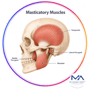
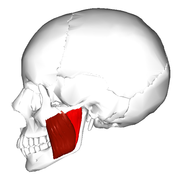
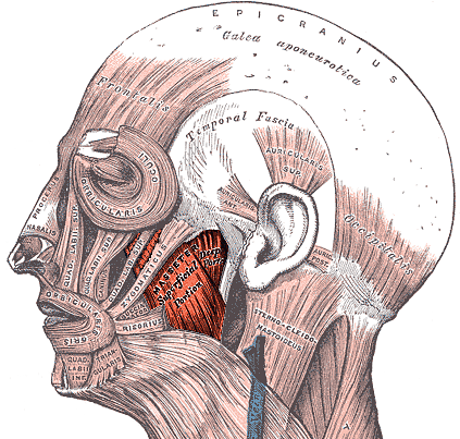
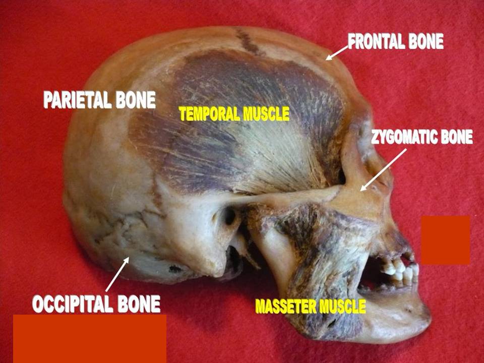
The muscle extends from the zygomatic arch of the viscerocranium to the lateral aspect of the mandible, forming the muscular bulk of the posterior cheek. Because of its short fibers, large physiological cross-sectional area, and favorable mechanical leverage relative to the temporomandibular joint (TMJ), the masseter is capable of generating extremely high mechanical forces.

Together with the medial pterygoid muscle, the masseter forms a muscular sling around the mandibular ramus, which stabilizes the mandible against the cranial base during mastication.

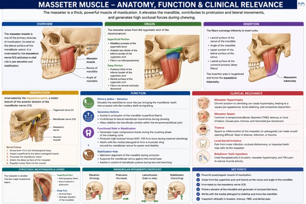
Structurally, the muscle is divided into two major anatomical layers:

Superficial part

Deep part

These layers differ in fiber orientation, attachment sites, and biomechanical contribution to mandibulr movement.

The masseter plays an essential role not only in jaw closure, but also in mandibular stabilization during occlusion, postural control of the mandible, and coordination of complex chewing movements.

ANATOMY

Origin

Superficial Portion

maxillary process of the zygomatic bone

anterior two-thirds of the inferior border of the zygomatic arch

The fibers of this portion run inferoposteriorly.

Deep Portion

posterior third of the inferior border of the zygomatic arch

medial surface of the zygomatic arch

The fibers of the deep portion run almost vertically downward toward the mandibular ramus

Exam Question

Explain the anatomical origin of the masseter muscle, distinguishing the superficial and deep portions, and analyze how the orientation of their fibers contributes to mandibular elevation, protrusion, and biomechanical stabilization during mastication.

Insertion

The fibers converge inferiorly to insert onto:

lateral surface of the mandibular ramus

angle of the mandible

upper portion of the lateral mandibular ramus

lateral surface of the coronoid process (deep fibers)

The insertion area is roughened and forms the masseteric tuberosity, reflecting the powerful traction forces generated by the muscle.

Exam Question

Describe the insertion of the masseter muscle onto the mandibular ramus and angle, including the formation of the masseteric tuberosity, and discuss how this attachment reflects the powerful occlusal forces generated during chewing.

Innervation

The masseter is innervated by the masseteric nerve, a motor branch of the anterior division of the mandibular nerve (cranial nerve V₃).

Course of the nerve:

arises from the mandibular nerve in the infratemporal fossa;

passes laterally across the lateral pterygoid muscle.

traverses the mandibular notch.

enters the deep surface of the masseter.

Exam Question

Trace the anatomical course of the masseteric nerve from the mandibular division of the trigeminal nerve (CN V₃) to the deep surface of the masseter muscle, and explain its functional significance in coordinated mastication.

FUNCTIONAL ROLE

Mandibular Elevation

The masseter is the primary elevator of the mandible. Contraction of its powerful vertical fibers elevates the mandibular ramus, producing jaw closure and bringing the mandibular teeth into precise occlusion with the maxillary teeth. This action is fundamental for biting, clenching, and the generation of high masticatory forces.

Exam Question

How does the anatomical architecture of the masseter enable it to function as the primary elevator of the mandible, and why is this action essential for effective occlusion, biting, and forceful jaw closure?

Jaw Movements

Masseter contributes to several accessory jaw movements. The superficial fibers assist in protrusion of the mandible, while unilateral contraction contributes to ipsilateral mandibular excursion during chewing. Continuous tonic activity also helps stabilize the mandibular condyle within the temporomandibular joint (TMJ), ensuring smooth and coordinated mandibular mechanics.

Exam Question

Explain how different fiber orientations within the masseter contribute to mandibular protrusion, lateral excursion, and dynamic stabilization of the temporomandibular joint during functional jaw movements.

Mastication

The masseter is the principal power muscle of mastication, generating the strong occlusal forces required during the crushing phase of chewing. Its large cross-sectional area enables efficient compression of food between the molar teeth. Functionally, it works synergistically with the medial pterygoid to form a muscular sling around the mandibular ramus, maximizing both chewing efficiency and force production.

Exam Question

Why is the masseter considered the principal power muscle of mastication, and how do its structural characteristics and synergistic relationship with the medial pterygoid optimize force generation during the chewing cycle?

TMJ Stabilization

Beyond movement generation, the masseter serves an important stabilizing function. It maintains mandibular alignment during occlusion, supports proper positioning of the mandibular condyle within the TMJ, and assists in controlling mandibular posture during both rest and clenching. This stabilizing activity contributes to balanced force distribution across the masticatory apparatus and promotes functional joint integrity.

Exam Question

Discuss the role of the masseter in maintaining mandibular alignment, condylar stability, and balanced force distribution within the temporomandibular joint during mastication, clenching, and mandibular posture control.

CLINICAL RELEVANCE

Masseter Hypertrophy

Occur in:

chronic bruxism (teeth grinding)

persistent jaw clenching

unilateral chewing habits

This can produce prominent mandibular angles and widening of the lower face.

Exam Question

Explain the anatomical and functional mechanisms underlying masseter hypertrophy, including the effects of chronic bruxism, persistent jaw clenching, and unilateral mastication on muscular enlargement, mandibular contour remodeling, and lower facial morphology.

Masseter Spasm

Occur in:

temporomandibular disorders (TMD)

tetanus infection

local dental irritation

This may lead to pain and restricted jaw movement.

Exam Question

Evaluate the pathophysiology and clinical significance of masseter muscle spasm, including its association with temporomandibular disorders, tetanus infection, dental irritation, and the resulting impairment of mandibular mobility and masticatory function.

Trismus Inflamation

May cause trismus, a condition characterized by difficulty opening the mouth.

causes include:

odontogenic infections

trauma

postoperative inflammation.


Exam Question

Discuss the anatomical and inflammatory mechanisms responsible for trismus involving the masseter muscle, including odontogenic infections, traumatic injury, and postoperative inflammation, and explain how these processes restrict mandibular opening and alter oral function.

Neurological Injury

Damage to

mandibular nerve (CN V₃) or its masseteric branch may cause:

weakness of jaw closure

reduced bite force

impaired mastication.

Exam Question

Analyze the clinical consequences of neurological injury affecting the mandibular nerve (CN V₃) or the masseteric nerve, including weakness of mandibular elevation, reduced bite force, impaired mastication, and disruption of coordinated masticatory biomechanics.

SUMMARY TABLE

Scroll to Top