Fontanells
Fontanelles are membranous fibrous gaps at the intersections of cranial sutures in the fetal and neonatal skull, representing regions of delayed intramembranous ossification. They function as dynamic osteogenic growth zones, permitting rapid postnatal brain expansion, facilitating cranial molding during birth, and maintaining mechanical compliance of the developing skull. These structures ensure the transition from a flexible neonatal cranium to a stable adult cranial vault.
CORE

Overview
Structurally, fontanelles consist of dense fibrous connective tissue derived from mesenchymal precursors, forming a continuous interface with the sutural ligaments that unite adjacent cranial bones.
Externally, they are covered by periosteum, while internally they are lined by dura mater, establishing a direct anatomical relationship between the cranial vault and intracranial structures.
These membranous regions occur at junctions where three or more bones meet, forming mechanically compliant yet structurally integrated zones within the skull.
The major fontanelles include 6 distinct regions: the unpaired anterior (bregmatic) and posterior (occipital) fontanelles, and the paired sphenoidal (anterolateral) and mastoid (posterolateral) fontanelles, each occupying characteristic positions within the developing cranial framework.
ANATOMY
Anatomical Location
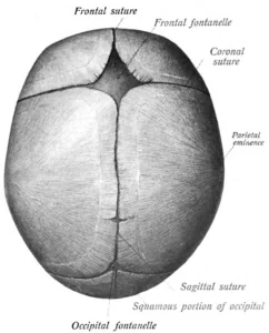
The anterior fontanelle is located at the junction of the frontal bone and the paired parietal bones, forming a diamond-shaped membranous area in the anterior cranial vault that corresponds to the adult landmark bregma. It represents the largest and most persistent fontanelle, typically closing between 18 and 24 months of age.
Posteriorly, the posterior fontanelle lies between the parietal bones and the occipital bone, forming a triangular region corresponding to the lambda, and it undergoes rapid closure within the first few months of life, usually by 2 to 3 months.
The sphenoidal, or anterolateral, fontanelle is situated at the convergence of the frontal, parietal, sphenoid, and temporal bones, corresponding to the adult landmark pterion, and it typically closes by approximately 6 months of age.
The mastoid, or posterolateral, fontanelle is located at the junction of the parietal, temporal, and occipital bones, corresponding to the asterion, and it persists longer, usually closing between 6 and 18 months. Through these relationships, fontanelles define critical transitional regions that later become key cranial landmarks used in anatomical orientation and clinical practice.
Exam Question
How do fontanelle junctions define cranial growth vectors and correspond to adult surgical landmarks.
Developmental Biology
Fontanelles arise from incomplete intramembranous ossification within the developing neurocranium, representing regions where mesenchymal tissue persists between adjacent ossification centers. During cranial development, bone formation proceeds centrifugally from primary centers within the cranial plates, while intervening zones remain unossified to accommodate growth.
Cranial expansion occurs predominantly at sutural margins, where osteogenic activity enables deposition of new bone and enlargement of the skull in a direction perpendicular to suture lines. This process is driven primarily by rapid cerebral growth and is regulated by molecular signaling pathways, particularly fibroblast growth factor (FGF) and transforming growth factor-β (TGF-β).
Functionally, fontanelles act as active osteogenic interfaces rather than passive gaps, coordinating cranial vault expansion and ensuring that skeletal growth remains synchronized with underlying brain development.
Exam Question
How do intramembranous ossification and sutural signaling regulate fontanelle persistence and cranial expansion.
Biomechanical Integration
Fontanelles, together with cranial sutures, form a compliant structural system that balances flexibility with stability.
In early postnatal life, rapid brain expansion – reaching the majority of adult volume within the first years – requires a skull capable of progressive enlargement without structural compromise.
During parturition, fontanelles permit transient deformation of the cranial vault, allowing overlapping of adjacent bones (molding) to facilitate passage through the birth canal. Following birth, this deformation reverses as intracranial pressures normalize.
Additionally, the fibrous architecture of fontanelles enables absorption and redistribution of mechanical forces across the cranial vault, reducing focal stress and protecting intracranial structures. Thus, they contribute critically to both developmental adaptability and mechanical resilience of the skull.
Exam Question
“How do fontanelles mediate viscoelastic deformation while preserving cranial structural integrity.”
Internal Reations
Internally, fontanelles are closely associated with the dura mater, which is firmly adherent to the inner surfaces of the developing cranial bones and extends beneath the membranous regions.
Beneath these areas, important vascular structures are situated, including the dural venous sinuses and meningeal vessels. In the posterior cranial region, the transverse sinuses lie in proximity to the posterior fontanelle, while in the lateral regions, the branches of the middle meningeal artery course beneath the sphenoidal fontanelle and adjacent sutural regions.
This close anatomical relationship between the membranous fontanelles, the dura mater, and intracranial vascular structures underlies their functional sensitivity to changes in intracranial pressure, as alterations in intracranial dynamics may be transmitted to the overlying soft tissues
Exam Question
“How do dura–vascular relationships at fontanelles explain their sensitivity to intracranial pressure dynamics.”
FUNCTIONAL ROLE
Neurocranial Expansion
Fontanelles function as osteogenic growth fields that permit cranial vault expansion in direct response to cerebral volumetric increase. Bone deposition occurs at adjacent sutural margins, allowing controlled enlargement perpendicular to suture lines while preserving structural continuity of the calvaria.
Parturition Adaptation
They provide a transient compliance system enabling biomechanical deformation of the neonatal skull during labor. Overlapping of cranial bones (molding) reduces biparietal diameter, facilitating passage through the birth canal without compromising intracranial structures
Mechanical Buffering
Fontanelles act as viscoelastic interfaces that dissipate and redistribute mechanical forces across multiple cranial bones, increasing energy absorption capacity and reducing localized stress transmission to the brain.
Architectural Coordination
They regulate spatial alignment and symmetry of cranial bones by guiding growth vectors, ensuring coordinated integration of the neurocranium and preventing distortion during rapid developmental expansion.
CLINICAL RELEVANCE
Intracranial Dynamics
Fontanelles reflect real-time intracranial pressure states due to their membranous nature: outward bulging indicates elevated pressure (e.g., hydrocephalus, meningitis), while depression signifies reduced intracranial volume (e.g., dehydration
Maturational Index
The sequence and timing of fontanelle closure serve as indicators of normal cranial ossification. Deviations suggest systemic or developmental pathology, including metabolic bone disease or endocrine disorders.
Growth Restriction
In craniosynostosis, premature suture fusion restricts growth perpendicular to the affected suture (Virchow’s law), forcing compensatory expansion in parallel planes and producing predictable cranial deformities.
Neurosonographic Window
Fontanelles provide acoustic windows for transfontanelle ultrasonography, enabling non-invasive visualization of ventricular size, intracranial hemorrhage, and parenchymal pathology in neonates.
SUMMARY TABLE
