Synchondrosis in general

Cartilaginous joints of the skull, known as synchondroses, are specialized primary cartilaginous articulations composed of hyaline cartilage that occur predominantly within the cranial base (chondrocranium). Unlike the cranial vault, which forms through intramembranous ossification, the bones of the cranial base originate from endochondral ossification, developing from a cartilaginous template during embryogenesis.

CORE

Overview

During early development, the cranial base exists as a continuous cartilaginous plate, which gradually differentiates into individual skeletal components separated by synchondroses. These cartilaginous joints function as active growth centers, analogous to the epiphyseal plates of long bones, permitting controlled elongation and angulation of the skull base during postnatal growth.

From an architectural perspective, the cranial base forms the central structural axis of the skull, supporting the brain and linking the neurocranium with the viscerocranium. Consequently, growth at synchondroses not only expands the cranial cavity but also determines the anteroposterior projection and spatial orientation of the facial skeleton.

ANATOMY

Overview

The spheno-occipital synchondrosis represents the principal postnatal growth center of the cranial base, forming a midline cartilaginous junction between the basisphenoid and basioccipital components of the chondrocranium. 

Functionally, it acts as a bidirectional endochondral growth plate, analogous to the epiphyseal plates of long bones, but uniquely oriented along the cranial base axis, thereby governing the longitudinal expansion and angular configuration of the skull base.

Exam Question

Why is the spheno-occipital synchondrosis considered the dominant postnatal growth center of the cranial base, and how does its midline position enable global craniofacial coordination?

Anatomical Position

It is located in the central cranial base, extending along the clivus between the dorsum sellae anteriorly and the anterior margin of the foramen magnum posteriorly

Superiorly, it supports the brainstem (pons and medulla) via the clival slope, while inferiorly it is structurally continuous with the craniovertebral junction, linking cranial base growth to axial skeletal alignment

Exam Question

How does its location along the clivus between the dorsum sellae and foramen magnum integrate brainstem support with cranial base growth and craniovertebral alignment?

Structural Organization

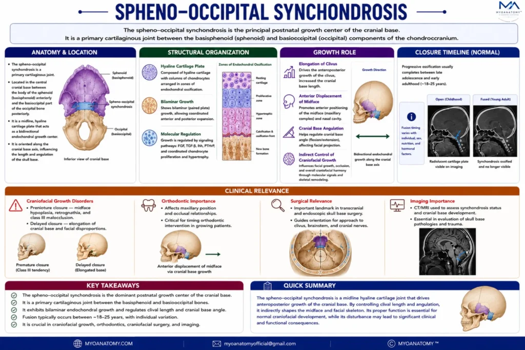
Histologically, the synchondrosis consists of paired plates of hyaline cartilage arranged symmetrically about the midline, each demonstrating classic zones of endochondral ossification:

resting cartilage

proliferative zone (chondrocyte column formation)

hypertrophic zone

calcification and ossification fronts

Unlike unidirectional long bone growth plates, this synchondrosis exhibits bilaminar growth, with proliferation occurring on both sides of the midline, producing bidirectional expansion. This results in coordinated anterior and posterior displacement of cranial base components.

Molecularly, growth is regulated by signaling pathways including FGF (fibroblast growth factor), TGF-β, Indian hedgehog (Ihh), and PTHrP, which coordinate chondrocyte proliferation, hypertrophy, and ossification timing.


Exam Question

How does the bilaminar arrangement of hyaline cartilage with mirrored endochondral zones generate bidirectional growth, and how does this differ functionally from long bone growth plates?

Growth Role

The spheno-occipital synchondrosis is the dominant determinant of cranial base length and angulation, driving:

elongation of the clivus

anterior displacement of the midface (maxillary complex)

spatial positioning of the nasal cavity and orbit

regulation of the cranial base angle (flexion/extension)

Through these mechanisms, it indirectly controls facial projection, occlusion, and overall craniofacial harmony, acting as a posterior growth driver within craniofacial growth models.

Exam Question

How does synchondrosis activity regulate cranial base length and angulation, and how do these changes mechanically drive anterior displacement of the midface?

Closure

The synchondrosis remains active throughout childhood and adolescence, undergoing gradual reduction in proliferative activity before complete ossification. 

Fusion typically occurs between 18–25 years, marking the cessation of cranial base growth and the formation of a continuous bony clivus.

Exam Question

What biological mechanisms explain its prolonged activity into adolescence and its fusion at 18–25 years as a marker of skeletal maturation?

Clinical Relevance

Premature fusion (synostosis) disrupts normal cranial base elongation, leading to:

midfacial hypoplasia (reduced anterior projection)

class III malocclusion (relative mandibular prognathism)

abnormal cranial base angle

craniofacial asymmetry

Conversely, delayed or excessive growth may alter facial proportions and occlusal relationships.

Radiologically, the synchondrosis is a key landmark in growth assessment, orthodontic planning, and forensic age estimation, as its fusion status correlates with skeletal maturity. 

In craniofacial surgery, it is critical for understanding deformity patterns and planning corrective interventions.

SUMMARY TABLE

Scroll to Top