Cartilaginous Joint
MYO CORE
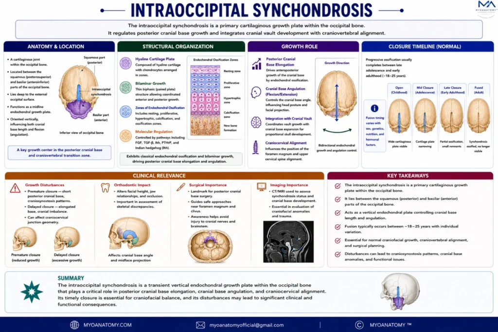
Intraoccipital Synchondrosis
The intraoccipital synchondroses are key regulators of posterior cranial base development, integrating occipital growth with foramen magnum formation and craniovertebral architecture.
OVERVIEW
Intraoccipital synchondroses are primary cartilaginous growth junctions within the occipital bone, representing the persistent interfaces between multiple embryonic ossification centers of the basioccipital, exoccipital, and supraoccipital components.
They function as transient but critical endochondral growth plates, coordinating the morphogenesis of the posterior cranial base and the craniovertebral transition zone.
Exam Question
Why are intraoccipital synchondroses considered transient but structurally critical growth interfaces, and how do they coordinate integration of the basioccipital, exoccipital, and supraoccipital components during posterior cranial base morphogenesis?
ANATOMY
Anatomical Position
These synchondroses are located within the posterior cranial base, surrounding the foramen magnum and extending across the basioccipital (anterior), exoccipital (lateral condylar), and supraoccipital (posterior squamous) regions.
They are positioned at the structural interface between:
the clivus anteriorly (continuous with spheno-occipital region)
the posterior cranial fossa superiorly (cerebellar support)
the craniovertebral junction inferiorly (atlas articulation)
Thus, they occupy a biomechanically strategic region linking neurocranium to axial skeleton.
Exam Question
How does their location around the foramen magnum and clival–craniovertebral interface position them as a biomechanical link between the posterior cranial fossa and axial skeleton?
Structural Organization
Histologically, intraoccipital synchondroses consist of hyaline cartilage plates undergoing endochondral ossification, with organized zones of:
proliferative chondrocytes aligned along regional growth vectors
hypertrophic expansion contributing to volumetric enlargement
vascular invasion and ossification fronts
Unlike a single dominant growth plate, these synchondroses act as a distributed growth network, coordinating multi-directional expansion of the occipital complex.
Their activity is regulated by molecular pathways including:
SOX9 (chondrogenic differentiation)
FGF signaling (growth modulation)
Indian hedgehog (IHH) (cartilage maturation and ossification timing)
Exam Question
How does their organization as multiple hyaline cartilage plates undergoing endochondral ossification function as a distributed growth network, rather than a single dominant growth center?
Growth Role
Intraoccipital synchondroses are essential for three-dimensional shaping of the posterior cranial base, contributing to:
foramen magnum expansion, ensuring adequate passage for the brainstem and vertebral arteries
development of occipital condyles, determining craniovertebral articulation geometry
formation and curvature of the clivus, influencing brainstem support and cranial base angulation
posterior cranial fossa volume, accommodating cerebellar growth
Functionally, they regulate posteroinferior cranial base growth, complementing anterior growth centers and ensuring balanced cranial base development along the sagittal axis.
Exam Question
How do intraoccipital synchondroses regulate foramen magnum expansion, occipital condyle formation, and clival curvature, thereby influencing brainstem accommodation and cranial base angulation?
Closure
These synchondroses ossify relatively early in postnatal development, with fusion occurring progressively as the occipital bone becomes a single continuous structure.
Their closure reflects the transition from:
→ growth phase (cartilaginous expansion)
→ structural stabilization (osseous integration)
After fusion, no further intrinsic growth occurs at these sites.
Exam Question
What is the significance of their early postnatal ossification, and how does this transition reflect the shift from cartilaginous growth to structural stabilization of the occipital base?
Clinical Relevance
Because intraoccipital synchondroses regulate the geometry of the craniovertebral junction, their disturbance has high clinical impact despite early closure.
Pathological alterations may lead to:
foramen magnum stenosis → brainstem compression
craniovertebral instability or malalignment → altered biomechanics between skull and spine
abnormal clival angle → brainstem kinking or posterior fossa crowding
occipital condyle dysmorphology → impaired atlanto-occipital articulation
These abnormalities are particularly relevant in:
skeletal dysplasias (e.g., achondroplasia)
Chiari malformations (posterior fossa underdevelopment)
craniovertebral junction anomalies
Radiologically, this region is critical in:
pediatric neuroimaging (posterior fossa development)
foramen magnum assessment
surgical planning for craniovertebral decompression
SUMMARY TABLE