Squamous Suture
The squamous (squamosal) suture is a lateral cranial vault articulation that unites the parietal bone with the squamous part of the temporal bone. Unlike the serrated sutures of the cranial vault, it exhibits a characteristic overlapping configuration, forming a smooth, curved junction along the lateral aspect of the skull. It contributes to the structural integrity of the lateral cranial wall and defines the superior boundary of the temporal fossa, a region intimately associated with the muscles of mastication.
CORE


Overview
The squamous suture extends in an arched course along the lateral surface of the skull, where the inferior margin of the parietal bone overlaps the superior margin of the squamous part of the temporal bone.
It is classified as a squamous-type fibrous joint, characterized by beveled, overlapping bone edges rather than interdigitating serrations.
This configuration allows the parietal bone to externally overlap the temporal bone, creating a mechanically efficient articulation adapted to withstand lateral compressive forces.
ANATOMY
Anatomical Location
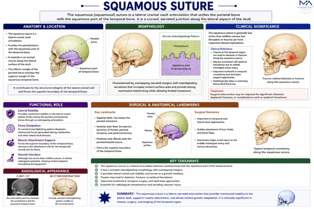
Superiorly, the squamous suture relates to the parietal bone, forming part of the lateral cranial vault, while inferiorly it overlies the squamous temporal bone, which contributes to the temporal fossa.
Anteriorly, it approaches the pterion, where it becomes continuous with the coronal and sphenoid articulations, and posteriorly it blends toward the parietomastoid region.
Through these relationships, it integrates the lateral cranial wall with both the cranial vault and the cranial base, forming a transitional zone between neurocranial and temporal regions.
Exam Question
Describe the anatomical location of the squamous suture, including the bones it connects and its role in forming the lateral cranial wall and temporal fossa.
Landmarks
Anteriorly, the squamous suture is closely associated with the pterion, a clinically significant landmark where the frontal, parietal, temporal, and sphenoid bones converge. This region overlies the anterior branch of the middle meningeal artery and is particularly vulnerable to traumatic injury.
Posteriorly, it approaches the asterion, marking the junction with the occipital and mastoid regions. These landmarks are essential for neurosurgical orientation, radiological interpretation, and surface anatomy localization of intracranial structures.
Exam Question
Identify the key landmarks associated with the squamous suture, particularly the pterion and asterion. Explain their anatomical and clinical significance.
Internal Relations
On the internal surface, the squamous suture corresponds to the lateral cranial wall, where the dura mater is adherent and vascular grooves for branches of the middle meningeal artery may be observed. This artery courses deep to the pterion and adjacent squamous region, supplying the dura mater.
The close relationship between bone, dura, and vascular structures renders this area clinically significant, as fractures involving the squamous suture may disrupt the middle meningeal artery, leading to epidural hematoma.
Exam Question
Outline the internal (endocranial) relations of the squamous suture, including its association with the dura mater and middle meningeal vessels. What are the clinical implications.
Sutures Features
The squamous suture is distinguished by its overlapping, beveled morphology, in contrast to the interdigitated patterns of serrate or denticulated sutures.
This structural arrangement allows slight gliding and enhanced resistance to lateral forces, particularly those generated by the temporalis muscle during mastication.
The fibrous connective tissue within the suture maintains a flexible interface while preserving stability, enabling both mechanical resilience and limited adaptive movement within the lateral cranial wall.
Exam Question
Explain the structural characteristics of the squamous suture. How does its overlapping morphology contribute to mechanical resilience and functional adaptation to masticatory forces.
FUNCTIONAL ROLE
Lateral Stability
The squamous suture provides essential mechanical stability to the lateral cranial wall by uniting the parietal and temporal bones through an overlapping articulation. This configuration reinforces the thin squamous temporal region, which would otherwise be structurally vulnerable.
The beveled arrangement distributes compressive forces across a broader surface, preventing focal stress concentration and contributing to the overall integrity of the cranial vault during both static loading and dynamic movement.
Force Dissipation
Due to its squamous (overlapping) morphology, this suture is particularly adapted to dissipate laterally directed mechanical forces. During mastication, powerful contractions of the temporalis muscle generate significant stresses along the temporal region.
The overlapping design allows controlled transmission and absorption of these forces, reducing the risk of structural failure and protecting underlying intracranial contents from repetitive mechanical strain
Muscle Adaption
The squamous suture plays a functional role in accommodating the biomechanical demands of the temporalis muscle, which occupies the temporal fossa directly inferior to the suture.
By forming the superior boundary of this region, it contributes to the attachment environment and force distribution of the muscle. This relationship ensures efficient transfer of muscular forces during chewing while maintaining the stability of the lateral skull.
Growth Interface
Although less active than midline sutures, the squamous suture retains osteogenic potential during development, allowing limited adaptive remodeling of the lateral cranial wall.
The fibrous connective tissue within the suture permits minor adjustments in bone positioning, facilitating coordinated cranial growth and maintaining proportional relationships between the cranial vault and temporal region
CLINICAL RELEVANCE
Pterion Fracture
The anterior portion of the squamous suture lies in close proximity to the pterion, a clinically critical weak point of the skull. Fractures in this region may rupture the anterior branch of the middle meningeal artery, resulting in an epidural hematoma.
This condition can rapidly increase intracranial pressure and requires urgent surgical intervention, making precise anatomical knowledge of this region essential
Epidural Hematoma
Due to its relationship with the middle meningeal vessels, trauma involving the squamous suture can lead to arterial bleeding between the dura mater and the inner table of the skull.
The accumulation of blood forms an epidural hematoma, which classically presents with a lucid interval followed by neurological deterioration. Early recognition based on anatomical landmarks is critical for life-saving management
Surgical Landmark
The squamous suture serves as an important reference in neurosurgical procedures involving the temporal region and lateral cranial vault. It helps guide surgical approaches to the middle cranial fossa and temporal lobe, allowing safe orientation relative to vascular and dural structures. Accurate identification minimizes the risk of intraoperative complications.
Radiological Differentiation
On imaging, the squamous suture must be distinguished from skull fractures, particularly in trauma cases. Its smooth, overlapping margins contrast with the sharp, linear appearance of fracture lines. Misinterpretation may lead to diagnostic errors; therefore, understanding its normal anatomical morphology is essential for accurate radiological assessment
SUMMARY TABLE
