Lamboid Suture
The lambdoid suture is a posterior cranial vault articulation that unites the paired parietal bones with the occipital bone, forming a curved, serrated junction across the posterior aspect of the skull. It represents a major structural boundary of the calvaria, separating the superior cranial vault from the posterior cranial region that overlies the cerebellum and occipital lobes.
CORE


Overview
The lambdoid suture extends transversely between the posteroinferior margins of the parietal bones and the superior border of the occipital squama, forming a configuration that resembles the Greek letter lambda (λ).
It constitutes a denticulated fibrous joint of the neurocranium, characterized by strong interdigitation between adjacent bones. As a primary posterior articulation, it stabilizes the occipital region while preserving a dynamic interface for cranial growth.
ANATOMY
Anatomical Location
Posteriorly, the lambdoid suture defines the articulation between the parietal bones and the occipital bone, marking the posterior limit of the parietal vault.
Superiorly, it connects with the sagittal suture, while inferolaterally it approaches the temporal region. Through these relationships, it contributes to the structural continuity of the cranial vault and the transition between superior and posterior cranial compartments.
Exam Question
Describe the anatomical position of the lambdoid suture, including the bones it unites and its role in defining the posterior boundary of the cranial vault.
Landmarks
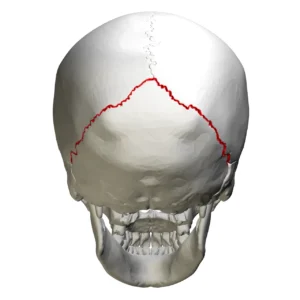
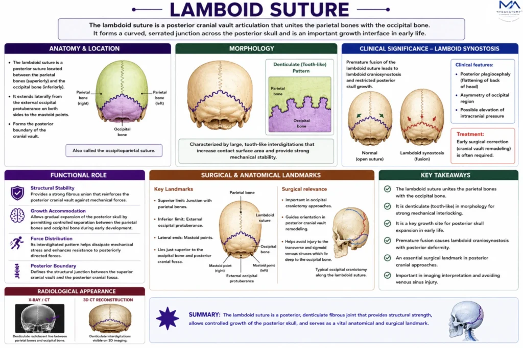
At its superior midpoint, the lambdoid suture intersects with the sagittal suture at the lambda, a key cranial landmark corresponding to the posterior fontanelle in early development.
Laterally, it extends toward the asterion, where the occipital, parietal, and temporal bones converge.
These landmarks are critical for anatomical orientation, radiological assessment, and neurosurgical access to the posterior cranial region
Exam Question
Identify the key landmarks associated with the lambdoid suture, particularly the lambda and asterion. Explain their anatomical and clinical importance.
Internal Relations
On the internal (endocranial) surface, the lambdoid suture is closely related to the grooves of the transverse sinuses, which lie along the attachment of the tentorium cerebelli.
The dura mater is firmly adherent in this region, and the adjacent bone often displays vascular impressions corresponding to venous sinuses and meningeal vessels.
This relationship is clinically significant, as injury in this region may compromise venous drainage of the posterior cranial fossa.
Exam Question
Outline the endocranial relations of the lambdoid suture, including its association with the transverse sinuses and dura mater. What are the clinical implications of this relationship.
Sutures Features
The lambdoid suture exhibits a denticulated (tooth-like) interdigitating pattern, characterized by larger and more pronounced bony projections compared to the finer serrations of the sagittal suture. This configuration increases the contact surface area and enhances mechanical resistance to posteriorly directed forces.
At the same time, the fibrous connective tissue within the suture permits limited flexibility and serves as an osteogenic interface, allowing controlled expansion of the posterior cranial vault during development.
Exam Question
Explain the structural morphology of the lambdoid suture. How does its denticulated interdigitating pattern contribute to mechanical stability and controlled cranial growth.
FUNCTIONAL ROLE
Posterior Expansion
The lambdoid suture functions as a key growth interface of the posterior cranial vault, permitting controlled expansion in response to the developing cerebellum and occipital lobes.
Osteogenic activity along the sutural margins allows gradual separation of the parietal and occipital bones, ensuring adequate accommodation of increasing intracranial volume during postnatal development.
Growth Regulation
Sutural patency is maintained through tightly regulated interactions between the sutural mesenchyme and the underlying dura mater, which provides molecular signals governing bone deposition and inhibition of premature fusion.
This coordinated signaling ensures balanced cranial morphogenesis and preserves the proportional development of the posterior cranial fossa.
Force Distribution
The denticulated architecture of the lambdoid suture enhances its ability to dissipate mechanical forces transmitted to the posterior skull.
By distributing stress across an interdigitated interface, it reduces localized strain and minimizes the risk of structural failure in regions subjected to external impact and muscular forces from the cervical attachments.
Structural Stability
The interlocking bony projections of the lambdoid suture provide significant mechanical reinforcement to the posterior cranial vault while maintaining limited flexibility.
This balance between rigidity and adaptability preserves cranial integrity, allowing the skull to withstand biomechanical stress while accommodating ongoing growth
CLINICAL RELEVANCE
Craniosynostosis Patterns
Premature fusion of the lambdoid suture results in lambdoid craniosynostosis, leading to restricted posterior cranial growth. This condition alters normal skull expansion dynamics and disrupts the balance between cranial vault compartments.
Plagiocephaly Deformity
Unilateral lambdoid synostosis produces posterior plagiocephaly, characterized by asymmetrical flattening of the occipital region with compensatory cranial distortion. Bilateral involvement may result in more global alterations in skull shape and volume distribution.
Surgical Lanmdmarks
The lambdoid suture serves as a critical anatomical guide in neurosurgical and craniofacial procedures, particularly through its relationship to the lambda and asterion. Accurate identification is essential for safe access to the posterior cranial fossa and avoidance of underlying venous sinuses
Radiological Differentiation
Radiologically, the lambdoid suture is distinguished by its irregular, interdigitated morphology, which contrasts with the linear appearance of skull fractures. This distinction is essential in trauma imaging to prevent misinterpretation and ensure accurate diagnosis
SUMMARY TABLE
