Coronal Suture

The coronal suture is a paired, serrated fibrous articulation of the cranial vault that forms the junction between the frontal bone anteriorly and the right and left parietal bones posteriorly. Oriented in the coronal plane, it extends transversely across the superior aspect of the skull from one pterion to the other, separating the anterior cranial vault from the parietal region. As a major component of the neurocranial sutural system, it serves both as a structural boundary and as a critical site of postnatal cranial growth.

CORE

Overview

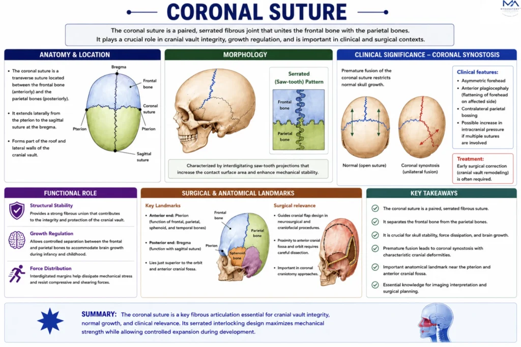
From an anatomical perspective, the coronal suture represents a complex interdigitating junction where adjacent cranial bones interlock through serrated margins. 

These irregular, tooth-like projections significantly increase the contact surface area between bones, thereby enhancing mechanical stability and resistance to shear forces. 


ANATOMY

Anatomical Relations

Anterior to the suture lies the frontal bone, contributing to the anterior cranial vault and the roof of the orbits, while posteriorly the parietal bones form the majority of the cranial vault.

Exam Question

Describe the anatomical relationships of the coronal suture, including the bones it unites and its role in separating the anterior and posterior cranial vault.

Landmarks

Superiorly, the coronal suture intersects with the sagittal suture at the bregma, a key cranial landmark, while laterally it approaches the pterion, where multiple bones converge

Exam Question

Identify the key landmarks of the coronal suture, particularly the bregma. Why is this junction clinically and surgically important?

Internal Relations

On the internal (endocranial) surface, the suture corresponds to the transition between frontal and parietal dural territories and is closely related to meningeal vascular grooves. 

Exam Question

Outline the endocranial relations of the coronal suture, including its association with dura and meningeal vessels. What are the clinical implications?

Sutures Features

The fibrous connective tissue within the suture, often referred to as the sutural ligament, maintains a dynamic interface that permits limited movement and growth while preserving structural integrity.

Exam Question

Explain the structural morphology of the coronal suture. How does its interdigitating pattern support stability and controlled growth?

FUNCTIONAL ROLE

Cranial Expansion

The coronal suture functions as a primary growth site of the cranial vault, permitting controlled separation of the frontal and parietal bones in response to progressive cerebral expansion. This is particularly critical for accommodating growth of the frontal lobes during early postnatal development.

Growth Regulation

Sutural activity is governed by tightly regulated interactions between the sutural mesenchyme and the underlying dura mater, which provides molecular signaling cues that maintain suture patency and coordinate osteogenic activity at the bone margins.

Force Dissipation

The serrated, interdigitating morphology of the coronal suture enhances its capacity to distribute and dissipate mechanical forces across the cranial vault, reducing focal stress concentration and limiting propagation of fracture lines during impact.

Structural Stability

By increasing the surface area of articulation between adjacent bones, the coronal suture provides significant biomechanical stability while preserving limited flexibility, ensuring both rigidity of the skull and protection of intracranial structures.

CLINICAL RELEVANCE

Coronal Synostosis

Premature fusion of the coronal suture disrupts normal cranial growth patterns, leading to craniosynostosis. Unilateral fusion produces anterior plagiocephaly, whereas bilateral fusion results in brachycephaly with reduced anteroposterior skull dimension.

Skull Deformity

Altered growth vectors secondary to suture fusion cause compensatory expansion in unaffected regions, producing characteristic craniofacial asymmetry and, in severe cases, functional impairment of orbital and cranial structures

Surgical Landmark

The coronal suture serves as a critical anatomical landmark in neurosurgical and craniofacial procedures, guiding orientation during craniotomy and facilitating safe access to the frontal lobes and anterior cranial fossa.

Radiological Assessment

On imaging, the coronal suture is essential for differentiating normal sutural anatomy from fractures and for diagnosing pathological fusion, with loss of the typical serrated pattern indicating synostosis

SUMMARY TABLE

Scroll to Top