Sagital Suture

The sagittal suture is a midline fibrous joint of the cranial vault that unites the right and left parietal bones. It forms the principal longitudinal articulation of the calvaria and serves as a key growth site, permitting transverse expansion of the skull in response to cerebral development. As a serrate suture, it combines mechanical stability with limited flexibility, maintaining cranial integrity while accommodating brain growth.

CORE

Overview

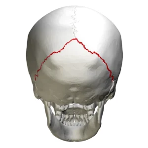
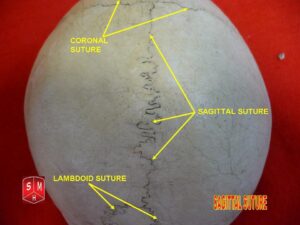
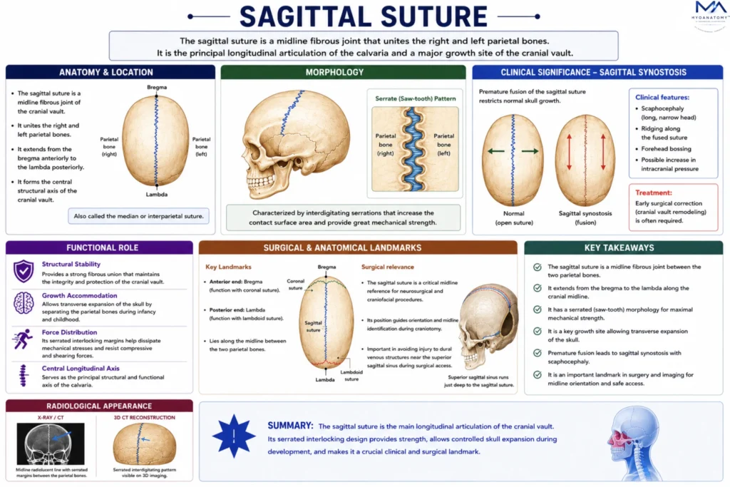
The sagittal suture extends along the median plane of the cranial vault, forming the central structural axis of the neurocranium. It runs between the two parietal bones from the bregma anteriorly to the lambda posteriorly, linking major cranial sutures and defining the longitudinal organization of the skull. 

Structurally, it consists of dense fibrous connective tissue interposed between interdigitating bone margins, creating a strong yet adaptable articulation. Internally, it corresponds to the groove for the superior sagittal sinus and is closely associated with dural attachments and meningeal vascular patterns, reflecting its importance in both structural and functional cranial architecture.

ANATOMY

Anatomical Location

Between the parietal bones, the sagittal suture forms the central longitudinal articulation of the cranial vault, separating the skull into right and left halves. 

Anteriorly, it meets the frontal bone at the bregma, while posteriorly it articulates with the occipital bone at the lambda, thereby defining the midline axis of the calvaria.

Exam Question

Describe the anatomical relations of the sagittal suture, including the bones it unites and its role in establishing the midline organization of the cranial vault.

Landmarks

Anteriorly, the sagittal suture intersects with the coronal suture at the bregma, a major developmental and clinical landmark. 

Posteriorly, it meets the lambdoid suture at the lambda, while along its course it corresponds externally to the vertex, the highest point of the skull in anatomical position.

Exam Question

Identify the principal landmarks of the sagittal suture, including the bregma and lambda. Why are these junctions important in clinical and developmental anatomy.

Internal Relations

On the internal (endocranial) surface, the sagittal suture corresponds closely to the course of the superior sagittal sinus, which lies within the attached margin of the falx cerebri. 

The adjacent inner table often shows grooves for meningeal vessels and arachnoid granulations, reflecting its intimate relationship with dural venous structures.

Exam Question

Outline the endocranial relations of the sagittal suture, particularly its association with the superior sagittal sinus and dural reflections. What are the clinical implications.

Sutures Features

The sagittal suture exhibits a serrate (interdigitating) morphology, characterized by complex, saw-tooth articulations between the parietal bones. 

This configuration increases surface area and mechanical stability, while maintaining a fibrous interface that permits controlled cranial expansion during growth without compromising structural integrity

Exam Question

Explain the structural morphology of the sagittal suture. How does its serrated interdigitation contribute to both mechanical strength and developmental adaptability.

FUNCTIONAL ROLE

Growth Accommodation

The sagittal suture serves as a primary site for transverse expansion of the cranial vault, allowing lateral separation of the parietal bones in response to the progressive enlargement of the cerebral hemispheres. 

This growth is mediated by osteogenic activity within the sutural margins and regulated by molecular signaling between the sutural mesenchyme and the underlying dura mater, ensuring coordinated cranial development.

Mechanical Stability

The interdigitating serrate architecture of the sagittal suture provides substantial mechanical strength, enabling the cranial vault to resist deformation under external forces. 

This structural arrangement minimizes displacement between adjacent bones while preserving the overall integrity of the skull during mechanical stress.


Force Distribution

Situated along the midline, the sagittal suture plays a critical role in distributing biomechanical forces symmetrically across both halves of the cranial vault. 

This reduces localized stress concentration and enhances the skull’s capacity to absorb and dissipate impact forces

Structural Alignemnt

The sagittal suture establishes the central longitudinal axis of the skull, maintaining bilateral symmetry and aligning the cranial vault with the underlying cerebral hemispheres. 

This alignment is essential for balanced cranial growth and proper spatial organization of intracranial structures

CLINICAL RELEVANCE

Sagittal Synostosis

Premature fusion of the sagittal suture leads to sagittal craniosynostosis, resulting in scaphocephaly, a characteristic elongated and narrow skull shape. This occurs due to restricted lateral expansion with compensatory growth in the anteroposterior dimension.

Intracranial Pressure

Early closure of the sagittal suture may impair normal cranial expansion, potentially leading to increased intracranial pressure. In severe cases, this can compromise cerebral development and result in neurological deficits.

Surgical Landmark

The sagittal suture serves as a critical midline reference in neurosurgical procedures. Its identification is essential for orientation and for avoiding injury to the superior sagittal sinus during surgical access to intracranial structures.


Radiological Assessment

On imaging, the sagittal suture provides an important diagnostic reference. Its serrated, interdigitated appearance allows differentiation from skull fractures, which typically present as linear, non-interlocking lines, and it is also assessed in evaluating pathological suture fusion

SUMMARY TABLE

Scroll to Top