Osteology
The orbital cavity (orbit) is a paired, pyramidal bony cavity of the skull that houses the eyeball (globe) and its associated muscular, vascular, and neural structures responsible for vision and ocular motility. It represents a major anatomical region where the neurocranium and viscerocranium converge, forming a complex skeletal framework that supports the visual apparatus and associated neurovascular pathways.
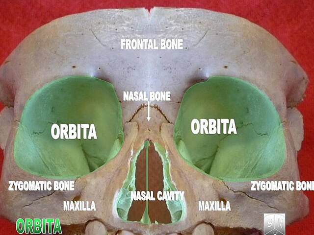
Each orbit provides mechanical protection, structural support, and spatial orientation for the globe while accommodating the extra-ocular muscles, optic nerve, ophthalmic vessels, lacrimal apparatus, and connective tissues that enable precise visual function.
CORE


Overview
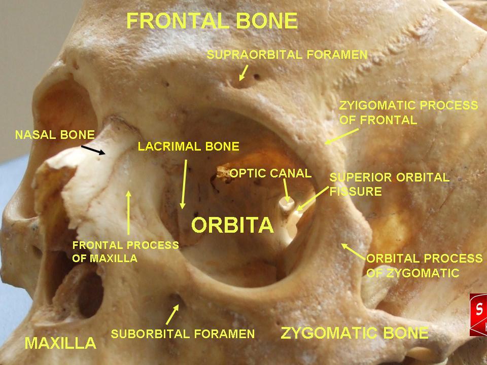
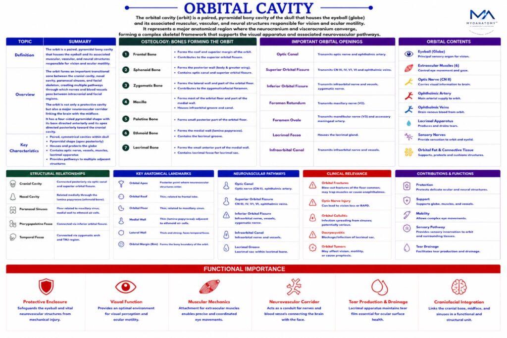
Anatomically, the orbit forms an important transitional zone between the cranial cavity, nasal cavity, paranasal sinuses, and facial skeleton, creating multiple pathways through which nerves and blood vessels pass between intracranial and facial regions.
The orbit therefore represents not only a protective cavity for the eye, but also a major neurovascular corridor linking the brain with the midface.
The orbit has a four-sided pyramidal shape, with its base directed anteriorly and its apex directed posteriorly toward the cranial cavity.
ANATOMY
Orbital Bones
The orbital cavity is formed by 7 bones of the skull, reflecting the complex integration between the neurocranial and facial skeleton.
These bones include:
frontal bone; sphenoid bone
zygomatic bone; maxilla
palatine bone; ethmoid bone
lacrimal bone
Together these bones form the 4 walls of the orbit, each with distinct structural properties and clinical significance.
Exam Question
Which seven bones constitute the orbit, and how does their composite arrangement reflect the structural integration between the neurocranium and viscerocranium.
Orbital Base
The orbital opening (base) is a quadrangular anterior aperture bounded by the orbital margin, formed by the
frontal /superior
zygomatic /lateral
maxilla /inferior
nasal /medial
Bones, providing structural protection and anterior access to the orbital contents.
Exam Question
Define the anatomical boundaries of the orbital base and explain its functional role in providing anterior access while maintaining structural protection of orbital contents.
Orbital Apex
The orbital apex lies posteriorly within the lesser wing of the sphenoid bone, where the orbit communicates with the middle cranial fossa through the optic canal and superior orbital fissure.
This region represents the point where major neurovascular structures enter the orbit, making it a critical anatomical landmark.
Exam Question
Describe the anatomical location of the orbital apex and analyze its significance as a neurovascular gateway between the orbit and middle cranial fossa.
Orbital Axis
The long axis of each orbit is directed posteromedially, meaning the two orbits converge toward the cranial cavity.
This orientation allows proper alignment of the visual axes, facilitating binocular vision and depth perception.
Exam Question
Explain the orientation of the orbital axis and evaluate its importance in binocular alignment and coordinated ocular function.
Superior Wall
The roof of the orbit is formed primarily by:
orbital plate of the frontal bone
lesser wing of the sphenoid bone
This wall separates the orbital cavity from the anterior cranial fossa, where the frontal lobes of the brain are located.
Important features of the orbital roof include the lacrimal fossa, which houses the lacrimal gland, responsible for tear production.
Because the orbital roof lies directly beneath the frontal lobe, traumatic injuries or fractures in this region may allow communication between the orbit and cranial cavity.
Exam Question
Describe the osteological composition of the orbital roof and discuss its anatomical relationship with the anterior cranial fossa and its clinical vulnerability in cranio-orbital trauma.
Inferior Wall
The floor of the orbit is formed mainly by:
the maxilla; the zygomatic bone
a small contribution from the palatine bone
This wall separates the orbital cavity from the maxillary sinus.
The floor contains the infraorbital groove and canal, which transmit the infraorbital nerve and vessels supplying the midface.
Because the orbital floor is relatively thin, it is particularly susceptible to blow-out fractures, where increased pressure within the orbit causes the floor to fracture downward into the maxillary sinus
Exam Question
Explain the structural composition of the orbital floor and analyze the anatomical basis and clinical mechanism of blow-out fractures.
Medial Wall
The medial wall is formed by: ethmoid bone (lamina papyracea):
lacrimal bone the frontal process of the maxilla
body of the sphenoid bone
The lamina papyracea of the ethmoid bone forms the largest portion of this wall and is extremely thin and delicate, separating the orbit from the ethmoidal air cells.
Because of this thin barrier, infections from the ethmoidal sinuses may easily spread into the orbit, leading to orbital cellulitis. The medial wall also contains the lacrimal groove, which contributes to the formation of the nasolacrimal canal.
Exam Question
Describe the components of the medial orbital wall and evaluate its clinical significance in the spread of infection from the ethmoidal air cells to the orbit.
Lateral Wall
The lateral wall is formed by:
zygomatic bone
greater wing of the sphenoid bone
This wall is the thickest and strongest wall of the orbit, providing significant protection for the globe against lateral trauma.
Its robust structure stabilizes the orbit and provides attachment surfaces for connective tissues and muscles associated with ocular movement.
Exam Question
Analyze the osteological structure of the lateral orbital wall and explain its role in providing maximal protection against lateral mechanical forces.
SUMMARY TABLE
