Openings
The orbit communicates with the cranial cavity and adjacent facial spaces through precisely positioned foramina and fissures that transmit neurovascular structures essential for vision, ocular motility, and orbital homeostasis.
CORE


Overview
The orbital openings are spatially organized at the apex and along the orbital walls, forming distinct pathways for the controlled passage of nerves and vessels.
This arrangement reflects a functional distribution where posterior openings relate to intracranial connections, while inferior and anterior pathways link the orbit to the face and deep facial spaces.
ANATOMY
Optic Canal
Located in the lesser wing of the sphenoid at the orbital apex, the optic canal provides a direct conduit between the orbit and the middle cranial fossa.
Transmits:
optic nerve (CN II)
ophthalmic artery
Functional significance:
primary afferent visual pathway and arterial supply to the orbit.
Clinical relevance:
compression → visual loss (e.g., optic canal fractures, tumors).
Exam Question
Explain the anatomical position of the optic canal within the sphenoid bone and analyze its functional significance in transmitting visual and vascular structures, including the clinical consequences of compressive or traumatic lesions at the orbital apex.
Superior Orbital Fissure
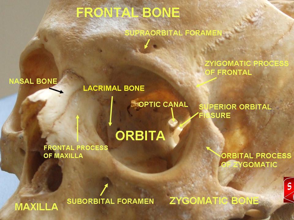
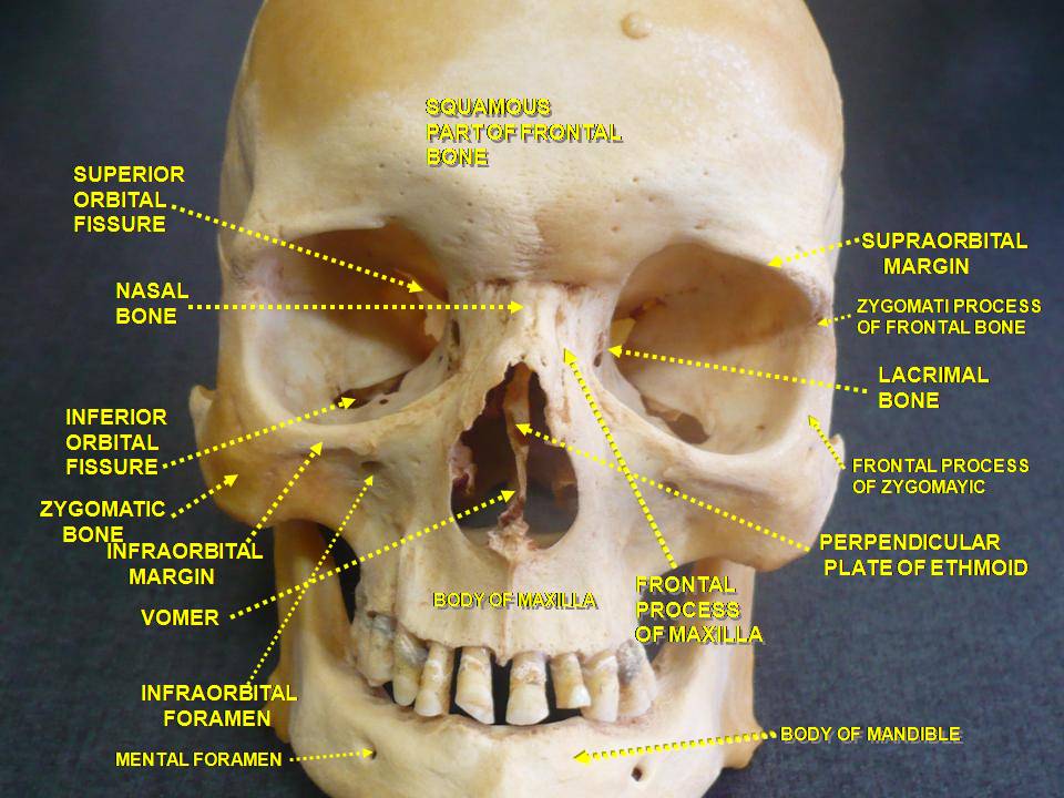
A slit-like opening between the greater and lesser wings of the sphenoid, connecting the orbit with the middle cranial fossa.
Transmits:
oculomotor nerve (CN III)
trochlear nerve (CN IV)
abducens nerve (CN VI)
ophthalmic nerve (CN V1: lacrimal, frontal, nasociliary branches)
superior ophthalmic vein
Functional significance:
controls extraocular movement and orbital sensation.
Clinical relevance:
lesions → ophthalmoplegia, ptosis, sensory loss (V1).
Exam Question
Describe the anatomical boundaries of the superior orbital fissure and evaluate its role as a major neurovascular conduit, detailing the cranial nerves transmitted and the resulting clinical deficits observed in lesions affecting this region.
Inferior Orbital Fissure
Located between the maxilla and greater wing of sphenoid, connecting the orbit with the pterygopalatine and infratemporal fossae.
Transmits:
infraorbital nerve (CN V2)
zygomatic nerve (CN V2)
infraorbital vessels
inferior ophthalmic vein (to pterygoid plexus)
Functional significance:
pathway for maxillary nerve distribution to midface.
Clinical relevance:
fractures → infraorbital numbness.
Exam Question
Discuss the anatomical relationships of the inferior orbital fissure with adjacent fossae and explain its role in transmitting maxillary nerve branches and vessels, including its clinical relevance in orbital floor fractures and midfacial sensory disturbances.
Nasolacrimal Canal
Extends from the lacrimal sac to the inferior nasal meatus.
Transmits:
nasolacrimal duct
Functional significance:
drains tears from orbit → nasal cavity.
Clinical relevance:
obstruction → epiphora (tear overflow).
Exam Question
Outline the anatomical course of the nasolacrimal canal and assess its functional role in tear drainage, including the pathophysiological basis and clinical presentation of nasolacrimal duct obstruction.
Infraorbital Canal & Groove
Located in the floor of the orbit (maxilla).
Transmits:
infraorbital nerve (CN V2)
infraorbital vessels
Functional significance:
supplies lower eyelid, cheek, upper lip.
Clinical relevance:
blow-out fractures → sensory deficit midface.
Exam Question
Describe the anatomical course of the infraorbital canal and groove within the orbital floor and analyze their role in transmitting neurovascular structures, including the clinical implications in blow-out fractures and sensory deficits of the midface.
SUMMARY TABLE
