
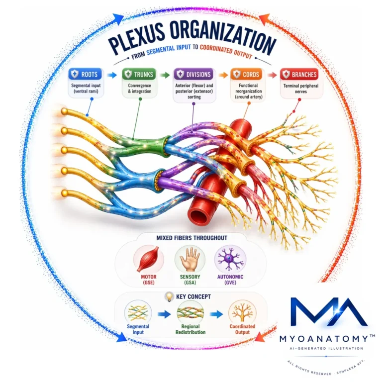
Plexus Organization
Peripheral nerve plexuses are neural redistribution networks formed by anterior (ventral) rami of spinal nerves, where axons converge, intermingle, and are reorganized into terminal peripheral nerves.
They exhibit a hierarchical structure (roots → trunks → divisions → cords → branches) that enables selective sorting of motor (GSE), sensory (GSA), and autonomic (GVE) fibers, ensuring each peripheral nerve contains multisegmental input.
Functionally, plexuses provide integrated innervation to muscles, joints, and skin, enabling:
Coordinated motor output (distributed motor unit recruitment)
Integrated sensory processing (proprioception, nociception, mechanoreception)
Reflex modulation and postural control
In contrast, posterior (dorsal) rami remain segmental, while anterior rami form plexuses supplying regions requiring complex, multi-joint neuromuscular coordination (limbs, anterior trunk).
Major plexuses:
Cervical (C1-C4) → neck, diaphragm; Brachial (C5-Th1) → upper limb
Lumbar (L1-L4) → anterior/medial thigh; Sacral (L4-S4) → posterior limb, foot
Overall, plexuses integrate motor, sensory, and autonomic pathways into unified systems supporting coordinated movement, adaptive biomechanics, and continuous sensorimotor feedback.
STRUCTURAL ORGANIZATION
Hierarchical organizationon
AI-Generated Illustration-MyoAnatomy
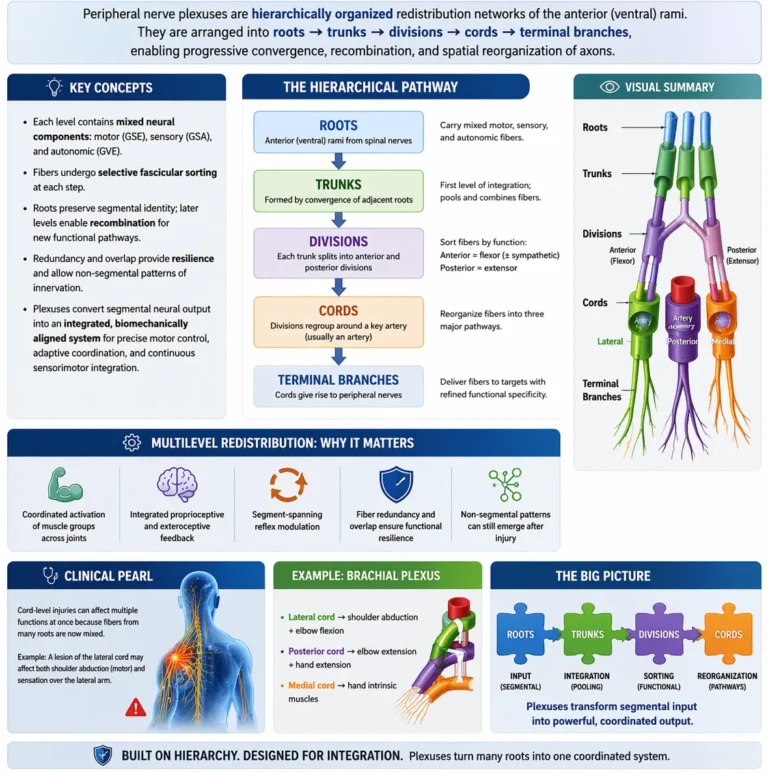
Peripheral nerve plexuses are hierarchically organized redistribution networks of the anterior (ventral) rami, arranged into roots → trunks → divisions → cords → terminal branches, enabling progressive convergence, recombination, and spatial reorganization of axons.
At each level, mixed neural components – motor (GSE), sensory (GSA), and autonomic (GVE)– undergo selective fascicular sorting, transforming initially segmental spinal input into functionally organized peripheral pathways. While roots preserve segmental identity, subsequent recombination permits target-specific fiber allocation according to modality and anatomical destination.
A key principle is division into anterior (flexor) and posterior (extensor) compartments, reflecting embryological patterning and ensuring biomechanical alignment of neural output with coordinated movement patterns.
This multilevel redistribution produces multisegmental innervation, allowing:
coordinated activation of muscle groups across joints
integrated proprioceptive and exteroceptive feedback
segment-spanning reflex modulation
Crucially, fiber redundancy and overlap ensure that each peripheral nerve contains contributions from multiple spinal segments, generating functional resilience, while also producing non-segmental patterns of deficit following plexus or root lesions.
Thus, plexuses convert segmental neural output into an integrated, biomechanically aligned system, enabling precise motor control, adaptive coordination, and continuous sensorimotor integration.
Exam Question
Explain how the hierarchical organization of peripheral nerve plexuses enables selective fiber redistribution, and critically analyze how this structural arrangement determines both functional integration and the pattern of neurological deficits following injury
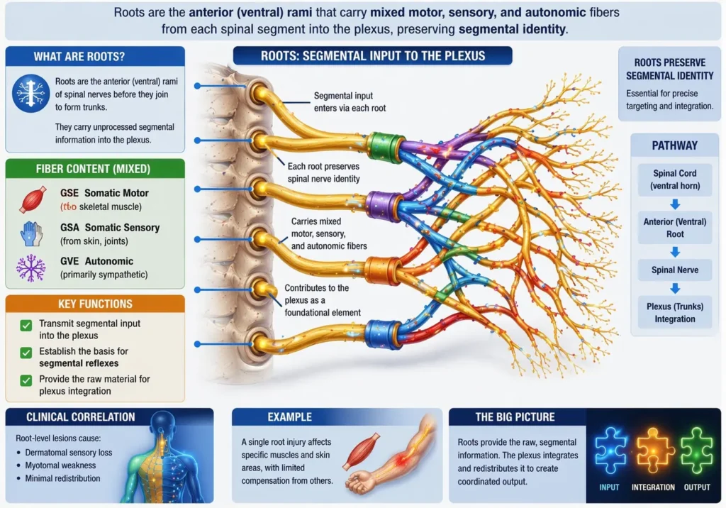
Roots
Roots (anterior/ventral rami) transmit mixed somatic motor (GSE), somatic sensory (GSA), and autonomic (GVE – primarily sympathetic) fibers from individual spinal segments. At this stage, segmental identity is preserved, maintaining strict correspondence with dermatomes and myotomes.
Functionally, roots represent the primary input streams into the plexus, carrying unprocessed segmental information. They establish the anatomical and physiological basis for segmental reflexes and radicular patterns of innervation.
Clinically, lesions at the root level produce dermatomal sensory loss and myotomal weakness, reflecting minimal redistribution.
AI-Genrated Illustration-MyoAnatomy
Exam Question
“Explain how preservation of segmental identity at the root level determines the clinical pattern of neurological deficits in radiculopathies.”
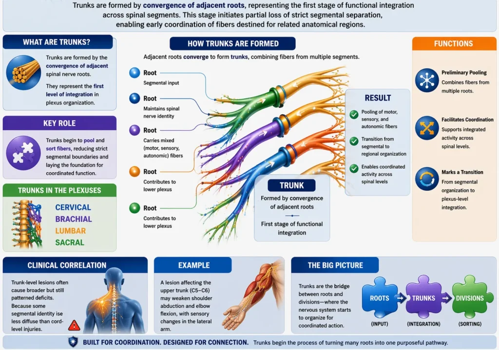
Trunks
Trunks are formed by convergence of adjacent roots, representing the first stage of functional integration across spinal segments. This stage initiates partial loss of strict segmental separation, enabling early coordination of fibers destined for related anatomical regions.
Functionally, trunks allow preliminary pooling of motor, sensory, and autonomic inputs, facilitating coordinated activity across multiple spinal levels. They mark the transition from segmental to regional organization.
Clinically, trunk lesions produce broader, mixed deficits, no longer confined to a single dermatome or myotome.
AI-Genrated Illustration-MyoAnatomy
Exam Question
“Critically analyze how the formation of trunks alters segmental organization and contributes to the emergence of regionally coordinated neural function.”
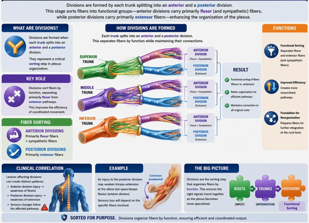
Devisions
Each trunk divides into anterior and posterior divisions, representing a critical step of functional sorting based on embryological and biomechanical principles.
Anterior divisions → flexor (ventral) compartments
Posterior divisions → extensor (dorsal) compartments
This segregation aligns neural output with movement patterns and compartmental muscle organization, ensuring efficient coordination of agonist groups.
Functionally, divisions represent task-oriented separation, organizing fibers according to functional biomechanics rather than segmental origin
AI-Genrated Illustration-MyoAnatomy
Exam Question
“Explain how the division of trunks into anterior and posterior components reflects embryological patterning and determines compartment-based motor organization.”
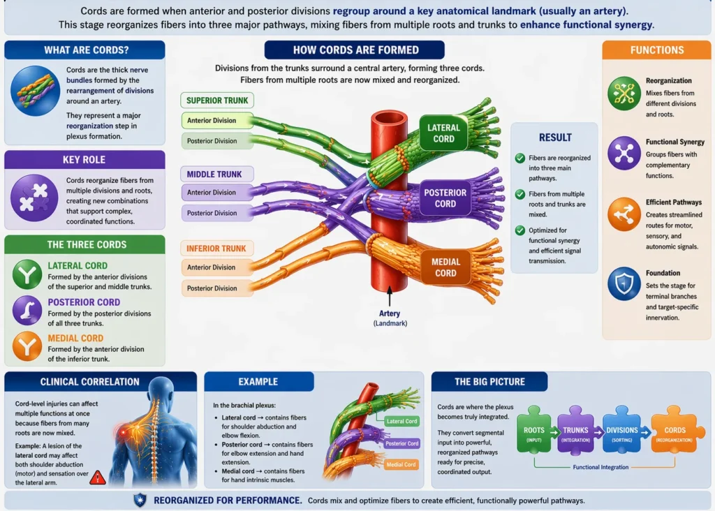
Cords
Divisions reorganize into cords, named relative to a central artery (e.g., axillary artery), where fibers undergo selective redistribution based on both functional role and target tissue.
At this level:
Fibers are no longer segmentally identifiable
They are functionally regrouped into integrated neurovascular pathways
Cords represent a higher-order integration stage, ensuring that peripheral nerves contain precisely calibrated mixtures of fibers required for coordinated muscle groups and joint systems.
Clinically, lesions produce complex, non-segmental deficits, reflecting disruption of integrated pathways.
AI-Genrated Illustration-MyoAnatomy
Exam Question
“Evaluate how fiber redistribution within cords contributes to functional integration, and explain why cord-level lesions produce non-segmental neurological deficits.”