
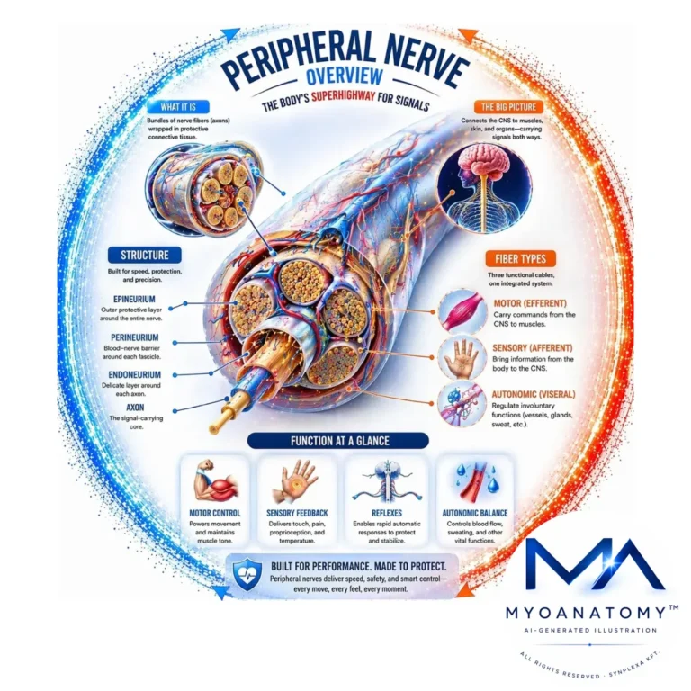
Peripheral Nerves
Peripheral nerves form the interface between the central nervous system (CNS) and peripheral tissues, enabling rapid bidirectional transmission for motor control, sensory processing, and autonomic regulation.
Consist of myelinated and unmyelinated axons organized into fascicles and supported by endoneurium, perineurium (blood–nerve barrier), and epineurium, providing both electrophysiological efficiency and mechanical protection. Schwann cells produce myelin with nodes of Ranvier, enabling saltatory conduction; conduction velocity depends on axon diameter and myelination. Fibers are classified into A (α, β, δ) and C types, reflecting functional specialization.
Functionally, peripheral nerves integrate motor (efferent), sensory (afferent), and autonomic fibers. At the neuromuscular junction, acetylcholine generates an end-plate potential, initiating muscle depolarization; force is regulated by motor unit recruitment and firing frequency.
Clinically, dysfunction arises from demyelination (reduced conduction velocity) or axonal injury (loss of transmission).
Overall, peripheral nerves operate as a dynamic system coordinating neural signaling with musculoskeletal function, ensuring precise and adaptive movement.
STRUCTURAL ORGANIZATION
Description
Graphical Abstract of a Peripheral Nerve” – Klimovich P., Rubina K., Sysoeva V., Semina E.; via Wikimedia Commons. Licensed under CC BY 4.0.
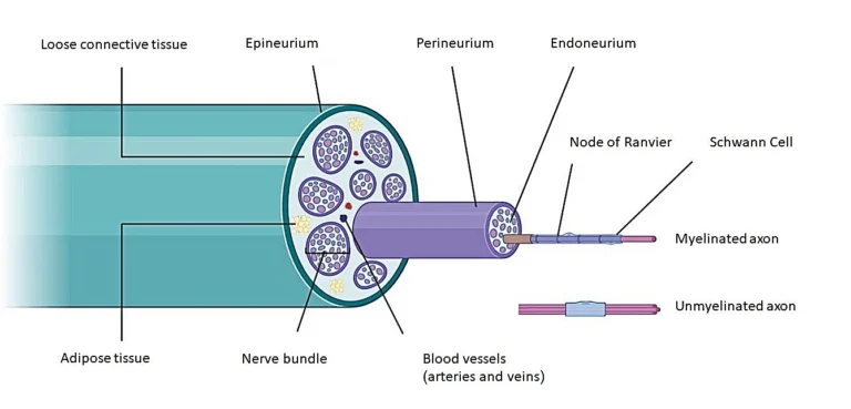
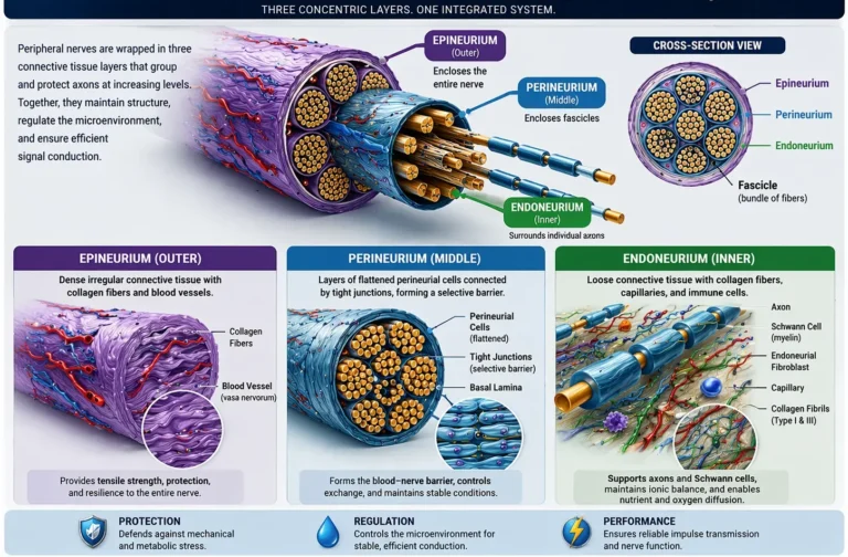
Peripheral nerves are hierarchically organized, compartmentalized structures in which axons are grouped into fascicles and supported by three concentric connective tissue sheaths – endoneurium, perineurium, and epineurium – that integrate structural, vascular, and electrochemical functions to preserve conduction fidelity under physiological and mechanical stress.
The endoneurium maintains the specialized microenvironment of individual axon–Schwann cell units, ensuring stable ionic gradients and extracellular composition required for membrane excitability and impulse propagation.
The perineurium forms a multilamellar diffusion barrier (blood – nerve barrier) that tightly regulates the exchange of ions and macromolecules, electrically isolates fascicles, and stabilizes the internal milieu necessary for consistent action potential conduction velocity.
The epineurium provides macroscopic structural integration, distributing tensile and compressive forces, permitting fascicular mobility, and protecting against mechanical deformation during limb movement.
This integrated system, supported by the vasa nervorum, sustains the high metabolic demands of axonal transport and ion pump activity; disruption of vascular supply or barrier integrity leads to impaired conduction and structural nerve injury.
Exam Question
Critically evaluate how the hierarchical organization of peripheral nerves – specifically the roles of the endoneurium, perineurium, and epineurium – maintains ionic homeostasis, mechanical resilience, and conduction velocity, and predict the functional consequences of disruption at each level.
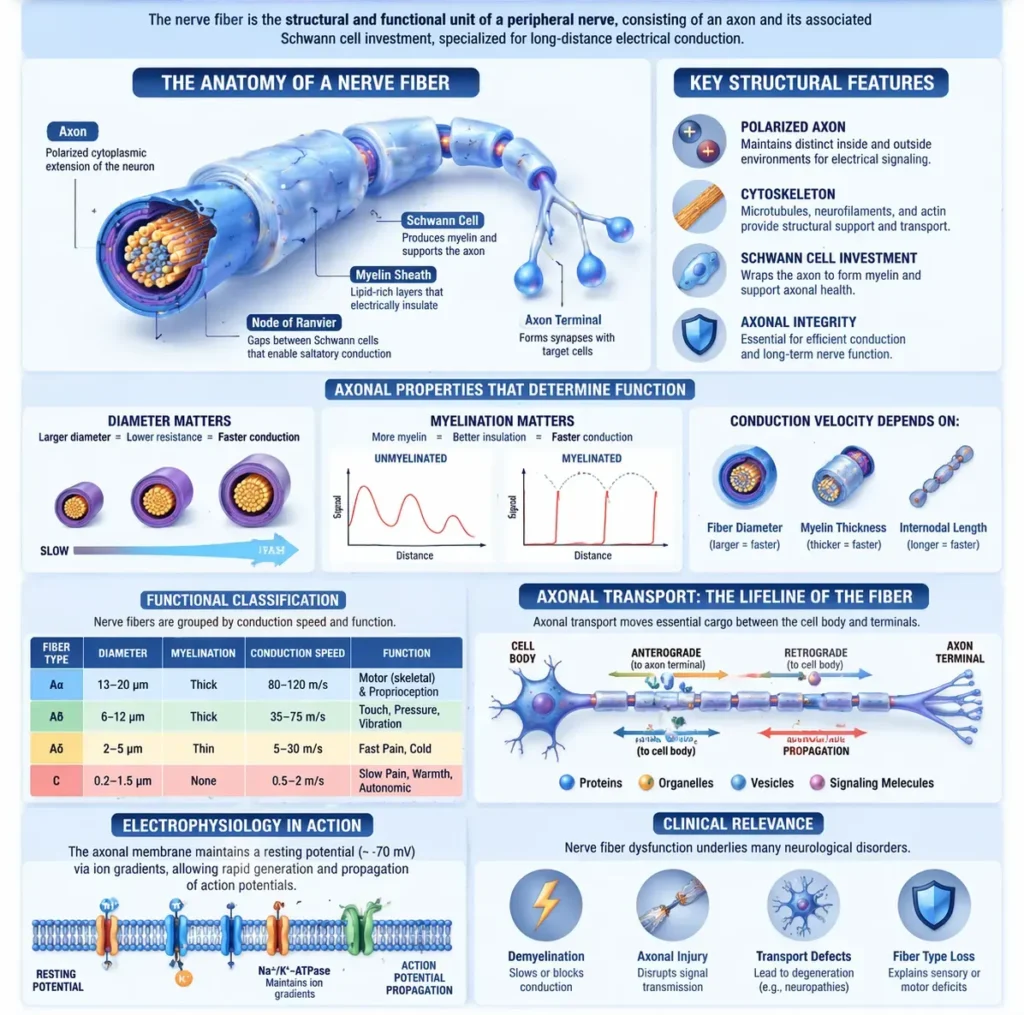
Nerve Fiber
The fundamental structural and functional unit of a peripheral nerve is the nerve fiber, consisting of an axon and its associated Schwann cell investment, specialized for long-distance electrical conduction. The axon is a polarized cytoplasmic extension of the neuron, containing a highly organized cytoskeleton (microtubules, neurofilaments, actin) that supports axonal transport and maintains structural integrity.
Axons exhibit marked variation in diameter, degree of myelination, and conduction velocity, which directly determine functional specialization. Large-diameter, heavily myelinated fibers conduct impulses rapidly via saltatory conduction, whereas small-diameter, unmyelinated fibers conduct more slowly via continuous propagation. Conduction velocity is therefore governed by fiber diameter, myelin thickness, and internodal length.
Functionally, nerve fibers are classified into A (α, β, δ) and C fibers, reflecting differences in conduction speed and modality (e.g., proprioception, motor control, fast vs slow pain). The integrity of the nerve fiber depends on axonal transport systems (anterograde and retrograde), which are essential for delivery of proteins, organelles, and signaling molecules between the cell body and peripheral terminals.
Electrophysiologically, the axonal membrane maintains a resting membrane potential through ion gradients established by Na⁺/K⁺-ATPase activity, and generates action potentials via voltage-gated ion channels. Disruption of axonal structure or myelin integrity impairs conduction, leading to functional deficits.
Thus, the nerve fiber represents a highly specialized unit integrating structural organization, metabolic support, and electrophysiological properties, enabling efficient and reliable neural transmission.
AI-Generated Illustration-MyoAnatomy
Exam Question
Critically analyze how axon diameter, myelination, and axonal transport mechanisms determine conduction velocity and functional specialization of nerve fibers, and explain the electrophysiological consequences of their disruption.
Myelinated Fibers
Myelinated nerve fibers are characterized by axons ensheathed by Schwann cells, which form a multilamellar myelin sheath composed of lipid-rich membranes that electrically insulate the axon. This sheath is organized into internodal segments separated by nodes of Ranvier, where the axonal membrane is exposed and enriched with voltage-gated Na⁺ channels.
Action potentials propagate via saltatory conduction, in which depolarization effectively “jumps” between nodes, significantly increasing conduction velocity while reducing metabolic demand. Conduction efficiency is determined by axon diameter, myelin thickness, and internodal length, optimizing rapid signal transmission in large A-type fibers responsible for motor control and high-fidelity sensory modalities.
Disruption of myelin integrity leads to conduction slowing or block, reflecting the critical role of myelin in maintaining electrical insulation and efficient impulse propagation
Exam Question
Explain the structural organization of myelinated nerve fibers and critically analyze how nodes of Ranvier and myelin architecture enable saltatory conduction and determine conduction velocity.
Unmyelinated Fibers
Unmyelinated nerve fibers lack a continuous myelin sheath, although axons are often partially enclosed within Schwann cell cytoplasm (Remak bundles). In these fibers, action potentials propagate via continuous conduction, requiring sequential depolarization along the entire axonal membrane, resulting in slower conduction velocities.
These fibers correspond primarily to C fibers, which are small-diameter and specialized for autonomic signaling, slow pain (nociception), and temperature transmission, where rapid conduction is not essential but sustained signaling is functionally advantageous.
Conduction velocity is limited by the absence of myelin and smaller axonal diameter, leading to increased capacitance and reduced membrane resistance compared to myelinated fibers.
Exam Question
Compare the structural and electrophysiological properties of unmyelinated and myelinated fibers, and explain how the absence of myelin influences conduction velocity, energy efficiency, and functional specialization
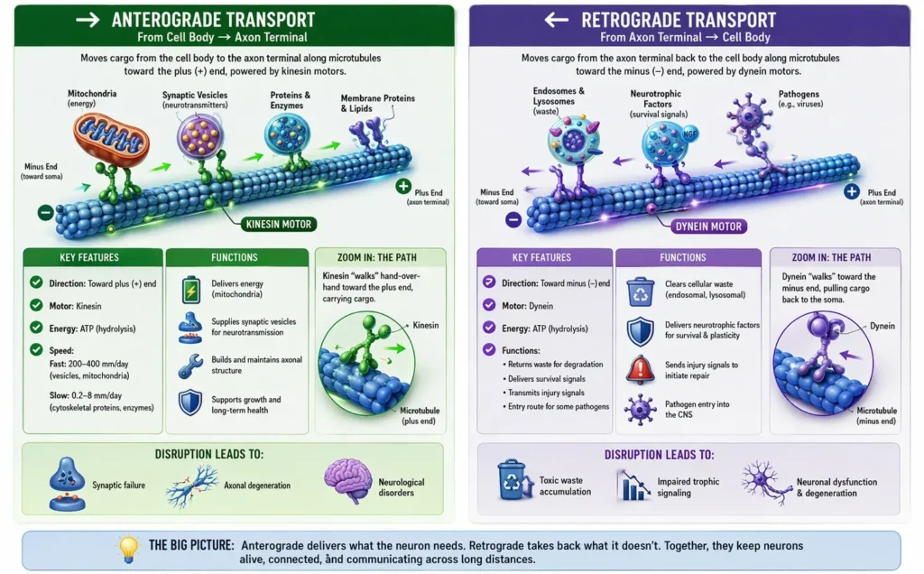
Axonal Transport
Axonal transport is a bidirectional, ATP-dependent system that moves organelles, proteins, and signaling complexes along polarized microtubules (plus-end distal, minus-end toward the soma), ensuring neuronal polarity, synaptic function, and long-distance cellular homeostasis.
Transport is mediated by motor proteins (kinesin, dynein) and regulated by intracellular signaling mechanisms.
AI-Generated Illustraton -MyoAnatomy
Anterograte Transport
Anterograde transport moves cargo from the cell body → axon terminal via kinesin motors toward the microtubule plus (+) end, powered by ATP hydrolysis.
Fast (≈200–400 mm/day): transport of membrane-bound cargo (mitochondria, synaptic vesicles, ion channels) via vesicular trafficking
Slow (≈0.2–8 mm/day): transport of cytoskeletal elements (tubulin, neurofilaments, actin) and soluble enzymes
Cargo binding occurs through adapter proteins, ensuring specificity of transport. Regulation involves phosphorylation states and Ca²⁺ signaling, modulating motor activity and cargo release.
Disruption leads to impaired synaptic delivery, axonal instability, and degeneration.
Exam Question
Explain the molecular mechanisms of kinesin-mediated anterograde transport, including microtubule polarity, ATP-dependent motor activity, and cargo regulation, and analyze its role in maintaining synaptic structure and function.
Retrograde Transport
Retrograde transport moves cargo from the axon terminal → cell body via dynein motors toward the microtubule minus (–) end, requiring the dynactin complex for cargo attachment and motor coordination.
Functions include:
Transport of endosomes, lysosomal degradation products
Delivery of neurotrophic factors (e.g., NGF) for survival signaling
Transmission of injury signals to initiate repair responses
This pathway is also exploited by pathogens (e.g., rabies virus, herpes simplex) for CNS entry.
Disruption impairs trophic signaling, leading to neuronal dysfunction and degeneration.
Exam Question
Critically evaluate the role of dynein – dynactin-mediated retrograde transport in neuronal survival and signaling, and discuss how its dysfunction contributes to neurodegenerative disease and pathogen propagation.
CONNECTIVE TISSUE LAYERS
Description
AI-Generated Illustration -MyoAnatomy
Peripheral nerves are supported by a system of concentric connective tissue sheaths that compartmentalize axons into structurally and functionally distinct units. These layers – endoneurium, perineurium, and epineurium – form an integrated framework that maintains mechanical integrity, regulates the intraneural microenvironment, and preserves the electrochemical conditions required for reliable impulse conduction.
Beyond structural support, this organization establishes controlled compartmentalization and selective permeability (via the blood–nerve barrier), ensuring stable ionic gradients, adequate vascular supply, and resistance to mechanical deformation during physiological movement.
Exam Question
Critically evaluate how the connective tissue organization of peripheral nerves establishes compartmentalization and selective permeability, and explain how these properties contribute to the maintenance of ionic homeostasis, conduction stability, and protection against mechanical and ischemic injury.
Endoneurium
The endoneurium is a specialized, delicate loose connective tissue that invests individual axon–Schwann cell units, forming the immediate microenvironment for nerve fibers. It is composed of longitudinally oriented type I and III collagen fibrils, endoneurial fibroblasts, resident macrophages, and a dense network of non-fenestrated capillaries with tight junctions, contributing to a controlled extracellular milieu. Each Schwann cell is enveloped by a basal lamina (rich in laminin and type IV collagen), which is critical for axonal support, regeneration, and guidance following injury.
Functionally, the endoneurium maintains a highly regulated endoneurial fluid compartment, ensuring stable ionic gradients and extracellular composition required for axonal excitability and impulse propagation. Its compliant structure allows limited deformation while protecting individual fibers from mechanical stress.
Exam Question
Analyze the cellular and extracellular composition of the endoneurium, and explain how its structural properties maintain the ionic microenvironment and regenerative capacity of peripheral nerve fibers.
Perineurium
The perineurium is a multilamellar, highly specialized connective tissue sheath that encloses nerve fascicles. It consists of concentric layers of flattened perineurial cells (modified myofibroblast-like cells) interconnected by tight junctions (zonula occludens) and supported by a basal lamina and collagenous matrix.
This structure forms the principal blood–nerve barrier, providing selective permeability to ions, macromolecules, and toxins, while maintaining osmotic and ionic stability within the fascicular compartment. It also serves as an electrical insulator, preventing current dissipation and preserving action potential propagation. The integrity of the perineurium is essential for maintaining conduction velocity and electrophysiological stability.
Exam Question
Critically evaluate the role of the perineurium as a blood–nerve barrier, and discuss how its structural organization contributes to electrical insulation, ionic regulation, and preservation of conduction velocity.
Epineurium
The epineurium is a dense irregular connective tissue forming the outermost sheath of peripheral nerves, composed of abundant type I collagen bundles, elastic fibers, fibroblasts, adipocytes, mast cells, and the larger vessels of the vasa nervorum. It organizes multiple fascicles into a cohesive nerve trunk and provides both structural support and mechanical adaptability.
Functionally, the epineurium confers tensile strength, allows fascicular mobility, and dissipates compressive, tensile, and shear forces encountered during limb movement. It also serves as a conduit for vascular supply, ensuring metabolic support to deeper nerve compartments. It is subdivided into interfascicular epineurium and external epineurium, enhancing both internal cohesion and external protection.
Exam Question
Explain the structural composition of the epineurium and analyze how it contributes to mechanical protection, vascular support, and functional integrity of peripheral nerves under dynamic physiological conditions.
Functional Significane
Peripheral nerves- hierarchical organization – ensures high-fidelity signal transmission
This architecture enables efficient propagation of action potentials by maintaining stable ionic gradients and electrical insulation, while simultaneously protecting nerve fibers from mechanical deformation, compression, and shear stress encountered during movement.
The integration of vascular supply (vasa nervorum) with connective tissue compartmentalization ensures continuous metabolic support, sustaining axonal transport, ion pump activity, and membrane excitability.
Functionally, this system allows peripheral nerves to coordinate motor output, sensory input, and autonomic regulation, enabling , and maintenance of homeostasis within the musculoskeletal system.
Exam Question
Critically evaluate how the structural organization of peripheral nerves integrates mechanical protection, vascular support, and electrochemical stability.
FUNCTIONAL CLASSIFICATION
Description
AI-Generated Illustration -MyoAnatomy
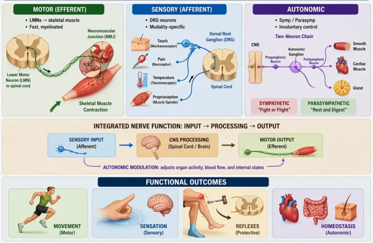
Peripheral nerves are functionally heterogeneous mixed structures composed of motor (efferent), sensory (afferent), and autonomic fibers, which together establish bidirectional communication between the central nervous system (CNS) and peripheral tissues. Within a single nerve trunk, these fiber types are organized into fascicles yet function as an integrated system, transmitting signals in opposite directions to coordinate neural activity.
Motor (efferent) fibers conduct impulses from the CNS to skeletal muscle, mediating voluntary contraction through neuromuscular transmission and precise motor unit recruitment.
Sensory (afferent) fibers convey information from peripheral receptors- including mechanoreceptors, nociceptors, and proprioceptors – to the CNS, enabling perception, reflex activity, and real-time modulation of movement.
Autonomic fibers regulate involuntary functions by controlling smooth muscle, cardiac activity, and glandular secretion, maintaining internal physiological homeostasis.
The integration of these components allows peripheral nerves to coordinate movement, sensation, and autonomic regulation, forming a dynamic system that supports reflex arcs, adaptive motor control, and continuous feedback between the body and central neural circuits.
Exam Question
Critically evaluate the functional classification of peripheral nerve fibers, and explain how the integration of motor, sensory, and autonomic components enables coordinated control of movement, perception, and homeostasis.
Motor Fiber
Motor (somatic efferent) fibers arise from lower motor neurons (LMNs) located in the anterior horn of the spinal cord or motor nuclei of the brainstem. These neurons are multipolar, and their axons exit via ventral roots, forming part of mixed peripheral nerves that project to skeletal muscle.
Motor axons are typically large-diameter, heavily myelinated Aα fibers, ensuring high conduction velocity required for rapid and precise movement. At the neuromuscular junction (NMJ), action potentials trigger Ca²⁺-dependent acetylcholine release, generating an end-plate potential that initiates muscle fiber depolarization and excitation–contraction coupling.
Functionally, motor output is organized into motor units, where:
α-motor neurons innervate extrafusal fibers → force generation
γ-motor neurons innervate intrafusal fibers → regulate muscle spindle sensitivity
This dual system enables alpha–gamma co-activation, maintaining muscle tone and allowing continuous proprioceptive feedback during contraction. Force modulation depends on:
Motor unit recruitment (spatial summation)
Firing frequency (rate coding)
Thus, motor fibers integrate central motor commands with peripheral biomechanical execution, forming the final common pathway for voluntary movement.
Exam Question
Critically analyze how motor fibers integrate lower motor neuron physiology, neuromuscular transmission, and motor unit organization, and explain how disruption at each level affects force generation, muscle tone, and movement precision.
Sensory Fiber
Sensory (afferent) fibers originate from pseudounipolar neurons in dorsal root ganglia (DRG), with a unique morphology allowing direct transmission from peripheral receptors to the CNS without synaptic interruption at the cell body. Their peripheral processes detect stimuli, while central processes enter the spinal cord via dorsal roots.
These fibers are functionally classified by diameter and myelination:
Aα (Ia, Ib) → proprioception (muscle spindle, Golgi tendon organ)
Aβ → fine touch, vibration
Aδ → fast pain, temperature
C fibers → slow pain, autonomic afferents
Sensory receptors transduce mechanical, thermal, or chemical stimuli into receptor potentials, which, if threshold is reached, generate action potentials. Proprioceptive input provides continuous feedback on muscle length, tension, and joint position, essential for:
Reflex arcs (e.g., stretch reflex)
Postural control
Cerebellar coordination
Sensory fibers thus form the afferent limb of neural circuits, enabling real-time modulation of motor output and adaptive movement control.
Exam Question
Evaluate the structural specialization of sensory neurons and fiber types, and explain how different modalities integrate to support proprioception, reflex activity, and coordinated motor control.
Autonomic Fiber
Autonomic (visceral efferent) fibers regulate involuntary functions via a two-neuron chain system consisting of preganglionic and postganglionic neurons. Preganglionic neurons originate in the intermediolateral cell column (sympathetic) or brainstem/craniosacral regions (parasympathetic), and synapse in autonomic ganglia.
Postganglionic fibers, typically unmyelinated C fibers, travel within peripheral nerves to innervate smooth muscle, cardiac muscle, and glands. In the limbs, autonomic fibers are predominantly sympathetic, regulating:
Vasomotor tone (arteriolar constriction/dilation)
Sudomotor activity (eccrine sweat glands)
Pilomotor responses (arrector pili muscles)
Neurotransmission involves:
Acetylcholine (preganglionic)
Noradrenaline (most postganglionic sympathetic fibers)
Functionally, autonomic fibers maintain local tissue perfusion, thermoregulation, and metabolic adaptation, dynamically adjusting blood flow in response to activity, temperature, and systemic demands.
Exam Question
Critically analyze the organization of autonomic fibers within peripheral nerves, including the preganglionic–postganglionic system and neurotransmitter mechanisms, and explain how this system regulates vascular tone, thermoregulation, and tissue homeostasis.
Functional Siginificance
Peripheral nerves function as integrated mixed neurovascular pathways containing motor(GSE), sensory (GSA), and autonomic (GVE) fibers, thereby establishing continuous bidirectional communication between the central nervous system and peripheral tissues. Their functional significance lies not simply in signal transmission, but in the coordinated regulation of movement, sensation, reflex control, vascular dynamics, and tissue homeostasis.
Motor fibers provide the efferent output required for skeletal muscle contraction, translating central neural commands into force production, posture, and coordinated movement.
Sensory fibers provide the afferent input necessary for touch, pain, temperature, and proprioception, supplying the CNS with continuous information about the mechanical and physiological state of tissues. This afferent feedback is essential for reflex integration, postural stability, and real-time modulation of motor activity.
Autonomic fibers, predominantly sympathetic within peripheral nerves, regulate involuntary functions such as vasomotor tone, sudomotor activity, and pilomotor responses, thereby controlling local perfusion, thermoregulation, and metabolic adaptation.
Functionally, these components operate as a unified system: sensory input informs central and spinal circuits, motor output executes adaptive responses, and autonomic activity maintains the internal conditions required for tissue function.
Exam Question
Critically evaluate the functional significance of motor, sensory, and autonomic fibers within peripheral nerves, and explain how their integration enables coordinated control of movement, reflex activity, vascular regulation, and tissue homeostasis
FUNCTIONAL IMPORTANCE
Motor Control
Peripheral nerves provide the efferent interface between the CNS and skeletal muscle, enabling controlled force generation via α-motor neuron activation and acetylcholine-mediated neuromuscular transmission. This initiates excitation–contraction coupling and mechanical force production.
Force output is precisely regulated through motor unit recruitment and rate coding, allowing graded contraction, coordinated movement, joint stabilization, and maintenance of posture under dynamic mechanical demands.
Sensory feedback
Peripheral nerves transmit continuous afferent input from mechanoreceptors, proprioceptors, and nociceptors to the CNS, encoding information on tissue deformation, position, and load. Muscle spindles detect changes in length and velocity, while Golgi tendon organs monitor tension.
This sensory input is integrated at spinal and supraspinal levels (including cerebellar circuits), enabling real-time modulation of motor output, ensuring accuracy, coordination, and adaptive control of movement.
Reflex Regulation
Peripheral nerves mediate spinal reflex circuits that generate rapid, automatic responses independent of conscious control. The stretch reflex maintains baseline muscle tone and postural stability, while withdrawal reflexes protect against noxious stimuli.
These reflexes operate through tightly regulated sensorimotor loops, allowing immediate neuromechanical adjustments and safeguarding tissues from excessive mechanical stress or injury.
Chronic Venous Insufficiency
Peripheral nerves function as integrated neurovascular units, coordinating motor output, sensory feedback, and autonomic regulation into a closed-loop system. This ensures efficient neuromechanical coupling, optimizing force transmission, balance, and movement adaptability.
Disruption of this integration leads to impaired excitation – contraction coupling, altered feedback control, and loss of coordinated movement, manifesting as weakness, instability, and inefficient motor patterns.stasis.
CLINICAL RELEVANCE
Peripheral Nerve Injury
Damage to motor fibers disrupts α-motor neuron output, resulting in reduced or absent muscle activation. This leads to weakness, paralysis, and impaired motor unit recruitment, compromising force generation and joint stability.
Chronic denervation produces muscle atrophy, decreased contractile efficiency, and altered fiber composition, leading to mechanically unstable and energetically inefficient movement patterns. Loss of coordinated activation further impairs fine motor control and posture.
Motor Disfunction
Damage to motor fibers disrupts alpha motor neuron output, resulting in reduced or absent muscle activation. This leads to weakness, paralysis, and impaired motor unit recruitment, ultimately compromising force generation and joint stability.
Prolonged denervation results in muscle atrophy and loss of neuromuscular efficiency, transforming movement into a mechanically unstable and energetically inefficient process.
Clinically, this is evident in conditions such as radial nerve injury (wrist drop), where loss of extensor function severely alters upper limb biomechanics.
Sensory Loss
Sensory fiber damage impairs transmission of afferent signals from mechanoreceptors, proprioceptors, and nociceptors, resulting in loss of proprioception, diminished tactile discrimination, and altered pain and temperature perception.
Without adequate sensory feedback, the CNS cannot effectively modulate motor output, leading to ataxia, impaired coordination, and instability. Aberrant signaling may produce neuropathic pain, characterized by spontaneous or exaggerated sensory responses due to dysfunctional afferent pathways.
Neuromechanical Disfunction
Peripheral nerve dysfunction represents failure of the integrated sensorimotor-autonomic system, disrupting coordination between motor output, sensory feedback, and reflex regulation.
This results in combined deficits in strength, coordination, timing, and adaptive control, impairing efficient movement and load distribution. The loss of neuromechanical coupling leads to compensatory patterns, instability, and progressive functional decline, particularly in conditions such as peripheral neuropathies.