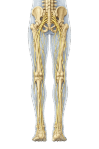
Neural Plexuses

Formation, Organization and Peripheral Innervation

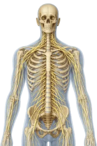
Neural plexuses are formed by the fusion of the ventral rami of adjacent spinal nerves, redistributing nerve fibers into peripheral nerves that provide coordinated motor, sensory, and autonomic innervation to the neck, trunk, and limbs.

Formation

Structure

Innervation

watermark

Cervical Plexus

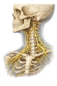
Fusion of the ventral rami of C1–C4 spinal nerves.

Forms superficial sensory and deep motor branches within the posterior cervical triangle.

Supplies the skin and muscles of the neck, diaphragm, and portions of the shoulder.

Injury may cause diaphragmatic paralysis (phrenic nerve) and sensory loss over the neck and shoulder.

Brachial Plexus

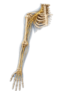
Fusion of the ventral rami of C5–Th1 spinal nerves.

Organized into roots, trunks, divisions, cords, and terminal branches.

Supplies motor and sensory innervation to the entire upper limb and shoulder girdle.

Trauma or birth injury may produce upper or lower limb weakness, sensory deficits, and characteristic plexopathies.

Lumbar

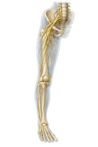
Fusion of the ventral rami of L1–L4 spinal nerves.

Forms major nerves within the posterior abdominal wall, including the femoral and obturator nerves.

Supplies the anterior and medial thigh, hip joint, and part of the lower abdominal wall.

Compression or injury may impair hip flexion, knee extension, and sensation over the anterior and medial thigh.

Sacral Plexus

Fusion of the ventral rami of L4–S4 spinal nerves.

Forms the sciatic nerve and major branches within the posterior pelvis.

Supplies the gluteal region, posterior thigh, most of the leg, and the entire foot.

Lesions commonly affect the sciatic nerve, causing lower limb weakness, sensory loss, and gait abnormalities.

Key Point

Only the ventral rami participate in neural plexus formation; the dorsal rami remain segmental and independently supply the intrinsic back muscles and overlying skin.

Scroll to Top