Joint Classification
MYO CORE
Structural Classification
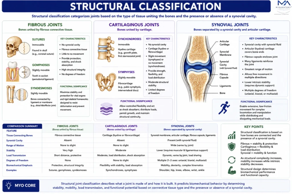
Joint structural classification organizes articulations according to the connective tissue uniting adjacent bones and the presence or absence of a synovial cavity. This structural organization fundamentally determines joint mobility, stability, load distribution, and biomechanical performance.
OVERVIEW
Structural joint classification categorizes joints according to the tissue connecting adjacent bones and the presence or absence of a synovial cavity. It provides the anatomical basis for understanding joint architecture, movement potential, mechanical stability, and load transmission throughout the skeleton.
Fibrous joints are united by dense connective tissue and prioritize stability; cartilaginous joints are connected by cartilage and provide controlled flexibility with shock absorption; synovial joints contain a synovial cavity and specialized articular structures that permit extensive and highly coordinated movement.
The structural composition of each joint determines its functional capacity, range of motion, resistance to mechanical stress, and role within kinetic chains. As structural complexity increases, mobility generally rises while intrinsic stability decreases, requiring greater ligamentous and muscular support.
Thus, structural joint classification forms the foundation for understanding joint biomechanics, skeletal integration, locomotion, posture, and the functional organization of the musculoskeletal system.
Exam Question
How does structural joint classification determine the functional capacity, mobility, stability, and biomechanical performance of a joint?
ANATOMY
Fibrous
Fibrous joints consist of bones united by dense fibrous connective tissue without the presence of a synovial cavity. These articulations exhibit minimal to no movement, with their primary function being mechanical stability, force transmission, and protection of adjacent structures. The rigidity of fibrous joints reflects their role in resisting tensile forces while maintaining structural integrity.
Three subtypes are recognized.
Sutures are immobile joints between cranial bones composed of short collagen fibers; they permit slight compliance during development and contribute to protection of the brain.
Syndesmoses involve bones connected by ligaments or interosseous membranes, allowing limited movement while maintaining stability and facilitating force distribution across adjacent bones.
Gomphoses are specialized peg-in-socket articulations between teeth and alveolar bone, stabilized by the periodontal ligament, permitting minimal movement to absorb and dissipate mechanical stress during mastication.
Exam Question
In a biomechanical context, how does the dense fibrous connective tissue architecture of fibrous joints determine their limited mobility, and how does this structural design contribute to force transmission, tensile resistance, and protection of vital structures?
Cartilaginous
Cartilaginous joints unite bones through cartilage,providing a balance between stability and limited mobility. These joints are specialized towithstand compressive forces, absorb mechanical stress, and permit controlled flexibility without the presence of a synovial cavity.
Two subtypes are distinguished.
Synchondroses are joints composed of hyaline cartilage, often temporary and associated with skeletal growth, such as the epiphyseal plates; they facilitate longitudinal bone growth and later ossify to form synostoses.
Symphyses consist of fibrocartilage and are characterized by high tensile strength and shock-absorbing capacity, as seen in intervertebral discs and the pubic symphysis; they permit slight movement while effectively resisting compression and shear forces.
Exam Question
How does the composition of cartilage (hyaline vs fibrocartilage) in cartilaginous joints determine their ability to resist compressive and shear forces, and how does this structural specialization support both skeletal growth and controlled mechanical flexibility?
Synovial
Synovial joints are characterized by the presence of a fluid-filled synovial cavity separating the articulating bones, enclosed within a fibrous capsule. This structural organization permits free movement (diarthrosis) and defines synovial joints as the principal sites of dynamic, multiaxial motion within the musculoskeletal system.
Their function is governed by specialized structural components.
Articular (hyaline) cartilage provides a smooth, low-friction surface that facilitates movement and distributes compressive loads across joint surfaces.
The synovial membrane produces synovial fluid, which reduces friction, nourishes avascular cartilage, and enhances movement efficiency through lubrication.
The fibrous capsule and associated ligaments confer mechanical stability while constraining and guiding motion within defined limits.
Periarticular structures, including tendons, bursae, and fibrocartilaginous elements such as menisci or labra, further optimize joint function by improving load distribution, reducing friction, and enhancing stability.
Synovial joints are additionally classified according to the shape of articulating surfacesand the axes of movement, including hinge, pivot, condyloid, saddle, and ball-and-socket types. These configurations determine the degrees of freedom, reflecting the joint’s specialized biomechanical role in coordinating movement within kinematic chains.
Exam Question
How do the structural components of synovial joints – particularly articular cartilage, synovial fluid, and capsuloligamentous structures – interact to regulate friction reduction, load distribution, and degrees of freedom, and how does this integration enable efficient multiaxial movement while maintaining joint stability?
SUMMARY TABLE