Cartilaginous Joint

MYO CORE

Overview

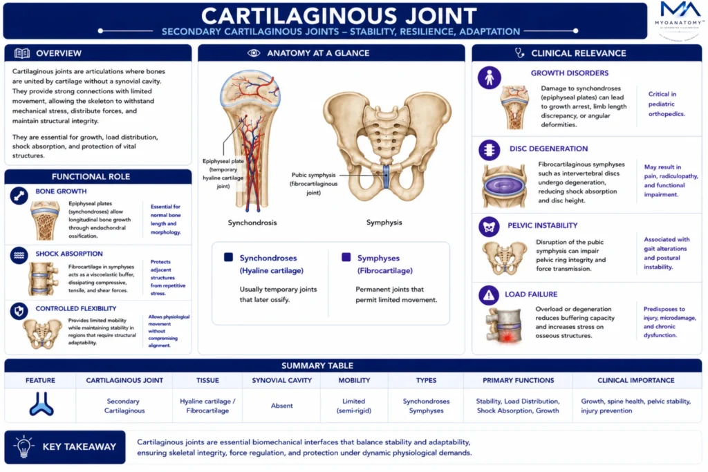
Cartilaginous joints are articulations in which adjacent bones are united by cartilage without a synovial cavity, forming continuous load-bearing interfaces that combine stability with controlled compliance. Structurally, they consist of hyaline cartilage, which resists compression via a hydrated proteoglycan-rich matrix, or fibrocartilage, which provides additional tensile and shear resistance through dense type I collagen.

OVERVIEW

Functionally, they permit limited, tightly regulated movement, preventing excessive displacement while adapting to mechanical loading. Their viscoelastic properties allow deformation under force with recovery, dissipating energy and reducing focal stress concentration. Biomechanically, they act as semi-rigid, energy-dissipating systems that distribute loads across skeletal segments, maintaining alignment and integrity under dynamic conditions, particularly within the axial skeleton.

They also play a key role in skeletal growth and remodeling, where cartilage functions as a transitional or permanent structural interface, optimizing durability and functional resilience under repetitive mechanical demand.

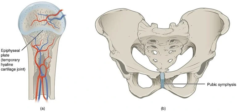
“Cartilaginous Joints” by OpenStax College, from Anatomy & Physiology, via Wikimedia Commons.  Licensed under CC BY 3.0

Why are cartilaginous joints considered essential biomechanical interfaces for force transmission, shock absorption, skeletal growth, and structural integrity within the human skeleton?

Exam Question

FUNCTIONAL ROLE

Bone Growth

Cartilaginous joints, particularly synchondroses, function as dynamic growth interfaces that enable longitudinal bone development through highly regulated endochondral ossification. Within the epiphyseal plate, spatially organized zones of chondrocyte proliferation, hypertrophy, and matrix mineralization generate progressive elongation while maintaining structural continuity.

This tightly controlled process determines final bone length and morphology, integrating cellular activity with mechanical loading conditions during growth.


Shock Absorption

Cartilaginous joints act as viscoelastic biomechanical buffers, particularly within the axial skeleton. Fibrocartilaginous structures, such as intervertebral discs, combine a tensile-resistant collagen network with a hydrated, proteoglycan-rich matrix that exhibits hydrostatic behavior under compression. 

This allows redistribution of forces across broader surfaces, reducing peak stress and protecting osseous structures from repetitive mechanical fatigue and microdamage.

Controlled Flexibility

These joints provide a precise balance between rigidity and mobility, permitting limited, controlled deformation while preserving structural stability. Their viscoelastic properties enable adaptive compliance under load, preventing excessive stiffness characteristic of fibrous joints and excessive mobility seen in synovial joints.

This controlled flexibility ensures maintenance of alignment and functional adaptability in weight-bearing regions under dynamic mechanical conditions.

Force Integration


Cartilaginous joints function as integrative mechanical interfaces that link adjacent skeletal segments into continuous load-bearing systems. By coordinating the transmission and distribution of forces across regions such as the vertebral column and pelvic ring, they ensure uniform stress dispersion and biomechanical coherence during movement, posture maintenance, and load-bearing activities. 

This integration minimizes localized overload and supports long-term structural integrity.

CLINICAL RELEVANCE

Growth Disorders

Damage to synchondroses, particularly the epiphyseal plates, disrupts normal endochondral ossification, leading to growth arrest, limb length discrepancies, or angular deformities.

These injuries are clinically critical in pediatric populations due to their long-term impact on skeletal development.

Disk Degeneration

Degeneration of fibrocartilaginous symphyses, such as intervertebral discs, is characterized by loss of proteoglycan content, reduced hydration of the nucleus pulposus, and structural disorganization of the annulus fibrosus. This leads to diminished hydrostatic pressure, decreased disc height, and compromised load distribution. 

Progressive weakening predisposes to annular tears and disc herniation, which may compress adjacent neural structures, resulting in pain, radiculopathy, and functional impairment.

Pelvic Instability

Alterations in the pubic symphysis, including ligamentous laxity, fibrocartilaginous degeneration, or mechanical disruption, can compromise the integrity of the pelvic ring. This leads to abnormal force transmission across the pelvis, particularly during dynamic loading conditions such as gait or pregnancy. 

Increased joint compliance results in instability, altered biomechanics, and impaired weight-bearing efficiency, with potential effects on posture and locomotion.

Load Failure

Compromise of cartilaginous joints disrupts their viscoelastic buffering capacity, leading to inefficient energy dissipation and increased stress concentration at adjacent osseous and articular structures. This promotes microdamage accumulation, accelerates degenerative processes, and predisposes to overuse injuries. 

The resulting loss of coordinated force transmission reduces overall biomechanical efficiency and contributes to progressive musculoskeletal dysfunction.

SUMMARY TABLE

Scroll to Top