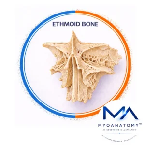
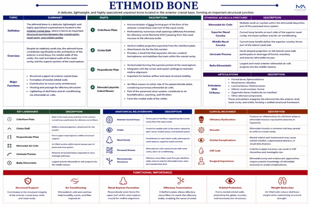
Ethmoid Bone

The ethmoid bone is a delicate, lightweight, and highly specialized unpaired bone located in the anterior cranial base, where it forms an important structural junction between the cranial cavity, nasal cavity, and orbital cavities. 

Despite its relatively small size, the ethmoid bone contributes significantly to the architecture of the anterior cranial fossa, the medial walls of the orbits, the roof and lateral walls of the nasal cavity, and the superior portion of the nasal septum.

CORE

Overview

Functionally, the ethmoid bone is closely associated with the olfactory system, as it transmits the olfactory nerve fibers (cranial nerve I) from the nasal cavity to the brain. It also contains the ethmoidal air cells, which form part of the paranasal sinus system and contribute to the conditioning of inspired air.

Because of its complex anatomy and numerous articulations with both cranial and facial bones, the ethmoid bone plays a critical role in maintaining the structural integration of the neurocranium and viscerocranium.

ANATOMY

Parts

Cribriform Plate

 A horizontal plate of bone that forms part of the floor of the anterior cranial fossa and the roof of the nasal cavity.

It is perforated by numerous small openings known as the olfactory foramina, through which the olfactory nerve filaments (CN I) pass from the nasal mucosa to the olfactory bulb located on the inferior surface of the frontal lobe.

This structure is therefore essential for the sense of smell (olfaction).

Crista Galli

Projecting superiorly from the midline of the cribriform plate is the crista galli, a vertical triangular projection.

The crista galli serves as an attachment site for the falx cerebri, a dural fold that separates the two cerebral hemispheres and stabilizes the brain within the cranial cavity.

Perpendicular Plate

The perpendicular plate extends inferiorly from the cribriform plate and forms the superior portion of the nasal septum, which divides the nasal cavity into right and left chambers.

Inferiorly, it articulates with the vomer and the septal cartilage, contributing to the structural framework of the nasal septum.

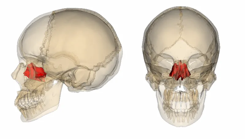
Ethmoidal Labyrinths (Lateral Masses)

On each side of the perpendicular plate lie the ethmoidal labyrinths, also known as the lateral masses.

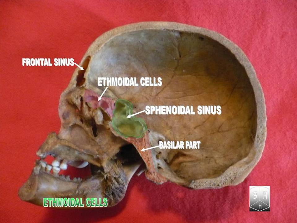
These structures contain numerous ethmoidal air cells, which form part of the paranasal sinus system. These air-filled cavities communicate with the nasal cavity and participate in humidifying, warming, and filtering inspired air.

The lateral surfaces of the labyrinths form the medial walls of the orbits, while their medial surfaces project into the nasal cavity.

Nasal Conchae

The medial surfaces of the ethmoidal labyrinths give rise to two curved bony projections:

superior nasal concha

middle nasal concha

These conchae increase the surface area of the nasal cavity, enhancing the processes of air filtration, warming, and humidification during respiration.

They also help regulate airflow patterns within the nasal cavity.

Exam Question

Explain how disruption of the cribriform plate–crista galli–ethmoidal labyrinth complex alters the functional interface between the central nervous system and the external environment, with emphasis on olfactory transmission and intracranial vulnerability.

Surfaces

The ethmoid bone contributes surfaces to several anatomical regions.

Cranial Surface –  superior surface of the cribriform plate forms part of the anterior cranial fossa, supporting the olfactory bulbs.

Orbital Surface –  lateral surface of the ethmoidal labyrinth forms the medial wall of the orbit. This thin plate of bone is known as the lamina papyracea because of its paper-like thinness.

Nasal Surface –  medial surface contributes to the lateral wall of the nasal cavity, supporting the superior and middle nasal conchae.

Exam Question

Critically analyze how the extreme thinness of the lamina papyracea influences the spread of infection between the ethmoidal sinuses and the orbit, and explain the anatomical basis of orbital complications.

Borders

The ethmoid bone articulates extensively with surrounding bones through its borders and processes.

The superior border articulates with the frontal bone, forming part of the anterior cranial base.

The posterior border articulates with the sphenoid bone, contributing to the cranial base.

The inferior borders articulate with bones forming the nasal septum and nasal cavity, including the vomer and maxillae.

Exam Question

Describe how the borders of the ethmoid bone contribute to anterior cranial base stability, and explain why fractures in this region preferentially result in CSF leakage rather than vascular compromise.

Articulations

The ethmoid bone articulates with 13 bones, reflecting its central position within the skull.

These include:

Frontal bone; Sphenoid bone

 Nasal bones; Maxillae

 Lacrimal bones; Palatine bones

 Inferior nasal conchae; Vomer

Through these articulations, the ethmoid integrates the cranial cavity, nasal cavity, and orbit into a unified structural framework.

Exam Question

Evaluate the role of the ethmoid bone as a structural “keystone” within the anterior skull, and explain how its multiple articulations facilitate coordinated integration of cranial, orbital, and nasal compartments

Landmarks

Cribriform plate  –  roof of the nasal cavity

Olfactory foramina –  passage for olfactory nerve fibers

Crista galli  –  attachment for falx cerebri

Perpendicular plate –  component of nasal septum

Superior and middle nasal conchae –  regulate airflow

Ethmoidal air cells  – part of paranasal sinus system

Lamina papyracea  – thin orbital wall

Exam Question

Discuss the clinical relevance of ethmoidal foramina and adjacent landmarks in relation to the transmission of olfactory fibers and the risk of intracranial infection.

FUNCTIONAL IMPORTANCE

Olfactory Transmission

The cribriform plate forms a highly specialized neuro-osteal interface, permitting passage of olfactory nerve filaments (CN I) from the nasal mucosa to the olfactory bulbs. 


This direct continuity represents one of the few sites where the central nervous system communicates with the external environment, making it both functionally critical and anatomically vulnerable.

Septal Integration

The perpendicular plate of the ethmoid constitutes the superior portion of the nasal septum, integrating with the vomer and septal cartilage to maintain midline alignment. 

This structural arrangement ensures laminar airflow, optimizing nasal conditioning functions including filtration, humidification, and thermal regulation

Orbital Positioning

The lamina papyracea forms the medial orbital wall, acting as a thin yet essential barrier between the orbit and ethmoidal sinuses. 

Despite its fragility, it maintains compartmental separation, preserving orbital integrity while allowing close anatomical and vascular relationships.

Pneumatic Modulation


The ethmoidal air cells contribute to skull pneumatization, reducing cranial weight while simultaneously enhancing respiratory air conditioning. 

Their mucosal lining supports humidification and contributes to vocal resonance through modulation of air-filled cavities

CLINICAL RELEVANCE

Barier Disruption

Fracture of the cribriform plate compromises the separation between the nasal cavity and anterior cranial fossa, leading to cerebrospinal fluid rhinorrhea. 

This creates a direct pathway for pathogens, predisposing to meningitis.

Neural Injury

Shearing forces at the cribriform plate can transect olfactory nerve filaments, resulting in anosmia. 

Due to the central connection of these fibers, recovery is often limited, making this a clinically significant and sometimes permanent deficit.

Orbital Compromise

The extreme thinness of the lamina papyracea facilitates rapid spread of infection from ethmoidal sinuses into the orbit. 

This may result in orbital cellulitis, subperiosteal abscess, or increased intraorbital pressure with risk to vision.

Sinus Propagation

Ethmoidal sinusitis may extend beyond local confines due to proximity to the orbit and anterior cranial fossa. 

Advanced spread can lead to intracranial complications such as meningitis, epidural abscess, or cavernous sinus involvement.

SUMMARY TABLE

Scroll to Top