Temporal Bone

The temporal bones are paired, highly complex bones forming the inferolateral walls of the skull and a significant portion of the cranial base. They lie between the sphenoid anteriorly, parietal superiorly, and occipital posteriorly, and play a central role in connecting the cranial vault with the facial skeleton and cranial base.

CORE

Overview

Each temporal bone houses the delicate organs of hearing and balance within its dense petrous portion, making it one of the most functionally specialized bones of the skull. In addition to protecting these sensory organs, the temporal bone forms part of the temporomandibular joint (TMJ), contributes to the cranial fossae, and serves as a conduit for several major cranial nerves and vascular structures.

Because of its structural complexity and numerous anatomical relationships, the temporal bone is of considerable importance in neurosurgery, otology, and craniofacial anatomy.

ANATOMY

Parts

Each temporal bone consists of 4 major anatomical parts.

These regions contribute to the cranial vault, cranial base, auditory system, and temporomandibular articulation.

Squamous Part

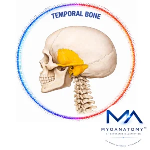
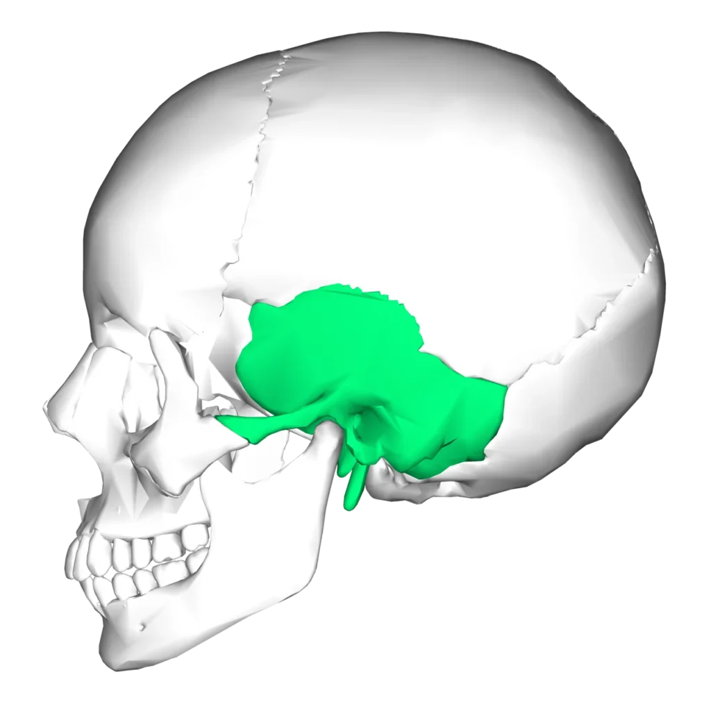
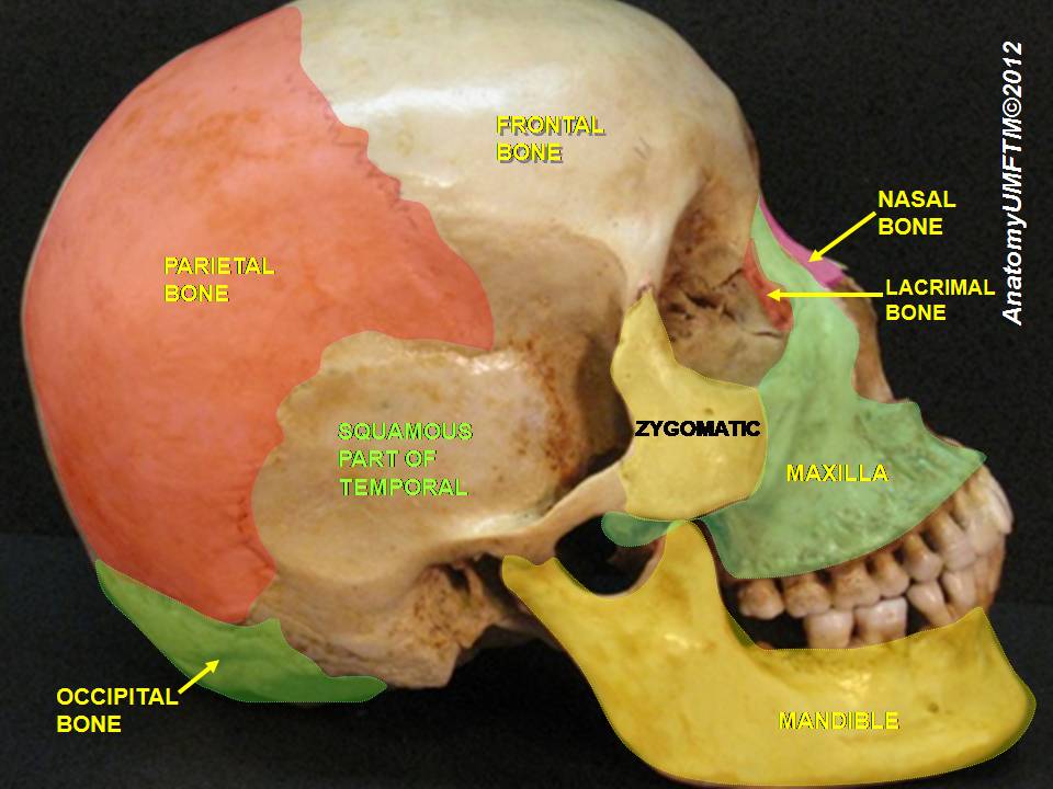
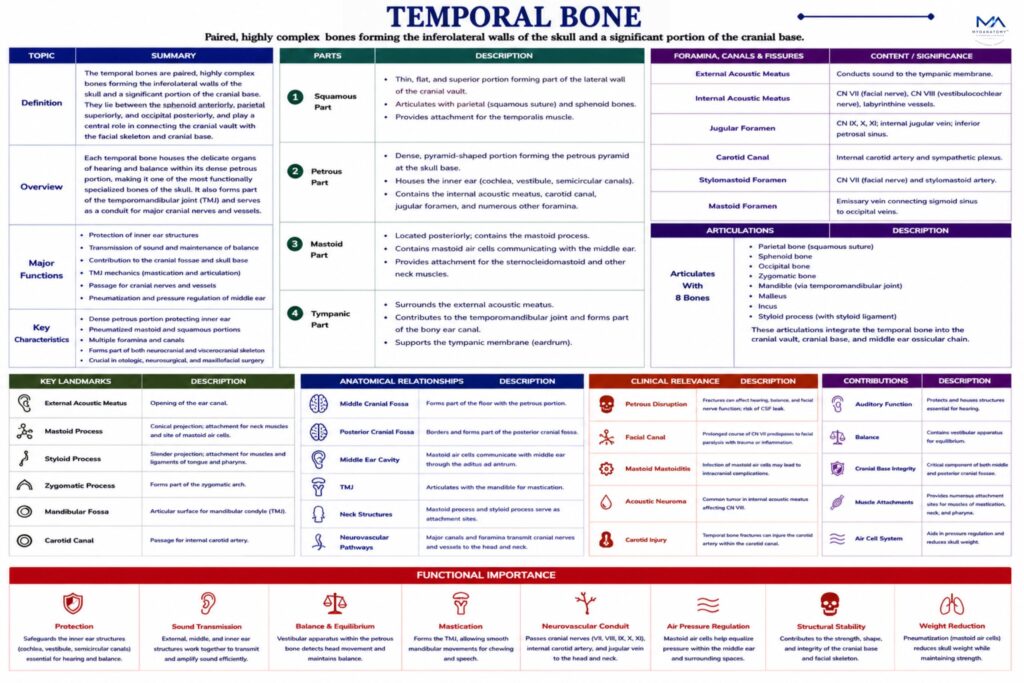
The squamous part is the thin, flat, and superior portion of the temporal bone that forms part of the lateral wall of he cranial vault.

Petrosus Part

The petrous part is a dense, pyramidal portion of the temporal bone situated at the base of the skull between the sphenoid and occipital bones. It is the hardest bone in the human body due to the protection required for the delicate structures it encloses.

Within the petrous part lie the structures of the inner ear, including:

cochlea (hearing), vestibule (balance),  semicircular canals (equilibrium)

major openings

Several important canals and foramina are located within the petrous portion:

Internal acoustic meatus- transmits the facial nerve (CN VII) and the vestibulocochlear nerve (CN VIII) to the inner ear.

Carotid canal-transmits the internal carotid artery, supplying blood to the brain.

Jugular fossa-forms part of the jugular foramen, through which pass:glossopharyngeal nerve (CN IX); vagus nerve (CN X); accessory nerve (CN XI); internal jugular vein

Mastoid Part

The mastoid part lies posterior to the external acoustic meatus and contains the mastoid process, a prominent conical projection that serves as an attachment site for several muscles involved in head movement, including the sternocleidomastoid muscle.

Within the mastoid process are numerous mastoid air cells, which communicate with the middle ear cavity and are lined with mucosa continuous with the middle ear.

These air-filled spaces contribute to

pressure regulation within the middle ear.

Tympanic Part

The tympanic part is a curved plate of bone that surrounds the external acoustic meatus, forming the bony portion of the ear canal.

This region supports the tympanic membrane (eardrum) and forms part of the boundary between the external and middle ear.

Exam Question

Explain how the squamous, petrous, mastoid, and tympanic parts of the temporal bone collectively integrate to support cranial protection, house neuro-sensory structures, and enable mandibular biomechanics.

Surfaces

The temporal bone contributes to several cranial surfaces:

Cranial Surface

Forms part of the middle cranial fossa, which accommodates the temporal lobes of the brain.

External Surface

Forms part of the lateral skull wall and supports muscular attachments and the external ear structures.

Inferior Surface

Forms part of the cranial base, containing openings for major neurovascular structures.

Exam Question

Correlate the cranial and external surfaces of the temporal bone with adjacent cranial fossae and extracranial regions, and explain how these relationships influence neurovascular protection and surgical access.

Borders

The temporal bone participates in several major cranial sutures:

Squamous border articulates with the parietal bone, forming the squamous suture.

Occipital border-articulates with the occipital bone, forming the occipitomastoid suture.

Sphenoidal borderarticulates with the greater wing of the sphenoid bone.

Exam Question

Analyze the role of temporal bone borders in forming major cranial sutures, and explain how these articulations contribute to mechanical force distribution across the skull.

Articulations

Each temporal bone articulates with 5 bones:

Parietal bone

Occipital bone

Sphenoid bone

Zygomatic bone

Mandible (via the temporomandibular joint)

These articulations integrate the temporal bone into both the neurocranial and facial skeletal framework.

Exam Question

Evaluate the articulations of the temporal bone with surrounding bones, and explain how these relationships enable temporomandibular joint function while maintaining cranial stability.

Landmarks

External acoustic meatus  opening of the ear canal

Mastoid process  – attachment for neck muscles

Styloid process  – slender projection serving as attachment for muscles and ligaments of the tongue and pharynx

Zygomatic process  – forms the zygomatic arch

Carotid canal  – passage for internal carotid artery

 Jugular fossa  – contributes to jugular foramen

Exam Question

Explain the anatomical and clinical significance of key temporal bone landmarks (e.g., external acoustic meatus, mastoid process, styloid process, carotid canal), particularly in relation to neurovascular transmission and pathology.

FUNCTIONAL IMPORTANCE

Neurosensory Housing

The petrous temporal bone forms a dense osseous capsule that mechanically isolates the cochlear and vestibular end-organs from external vibrational and pressure disturbances.

This structural rigidity preserves fluid dynamics of endolymph/perilymph, enabling precise mechanoelectrical transduction required for auditory frequency discrimination and vestibular spatial orientation.

Cranial Transmission

The temporal bone functions as a neurovascular gateway, transmitting critical structures (CN VII, VIII, internal carotid artery) through tightly constrained canals.

Its architecture ensures spatial segregation and protection of high-risk pathways while maintaining low-resistance continuity between intracranial and extracranial compartments, essential for uninterrupted neural signaling and cerebral perfusion

Mandibular Mechnaics

Through formation of the temporomandibular joint, the temporal bone enables biaxial joint mechanics combining rotation (hinge) and translation (gliding).

This allows dynamic modulation of occlusion, mastication force vectors, speech articulation, and airway patency, integrating skeletal movement with neuromuscular control.

Musculoskeletal Anchoring

The mastoid and styloid processes act as lever arms for muscle attachment, optimizing force transmission for head posture, cervical rotation, and deglutition mechanics.

These structures convert muscular contraction into controlled multidirectional movements, stabilizing the skull while coordinating cranio-cervical and pharyngeal functional units.

CLINICAL RELEVANCE

Petrosus Disruption

Fractures of the petrous temporal bone compromise the otic capsule and intratemporal neurovascular pathways, directly affecting the cochlear and vestibular apparatus.

This results in sensorineural hearing loss and vertigo, often accompanied by facial nerve paralysis due to its confined course within the facial canal.

Associated dural injury may produce CSF otorrhea, reflecting pathological communication between the cranial cavity and middle ear.

Facial Canal

The facial nerve’s prolonged intratemporal course within a rigid bony canal renders it highly susceptible to compression from inflammation or edema.

Even minimal swelling can produce complete ipsilateral facial paralysis, with additional deficits in lacrimation, salivation, and taste, depending on the level of involvement.

This highlights the clinical importance of anatomical confinement in amplifying neural dysfunction

Mastoid Continuity

The mastoid air cell system forms a continuous pneumatic extension of the middle ear, allowing infection to propagate beyond its initial site.

Spread may involve the sigmoid sinus (thrombosis) or intracranial structures, leading to meningitis or abscess formation.

These complications arise due to thin bony partitions and direct anatomical continuity between extracranial and intracranial compartments.

Acoustic Compression

Lesions within the internal acoustic meatus (e.g., vestibular schwannoma) compress CN VII and CN VIII within a confined osseous canal.

This produces a characteristic progression from sensorineural hearing loss and balance disturbance to facial nerve dysfunction, reflecting the topographic arrangement and limited space for expansion.

SUMMARY TABLE

Scroll to Top