Sphenoid Bone

The sphenoid bone is a complex, unpaired bone situated centrally at the base of the skull, forming a critical structural component of the cranial base. Because it articulates with all other bones of the neurocranium and several bones of the viscerocranium, it is often described as the “keystone” of the cranial floor.


CORE

Overview

The sphenoid contributes to the formation of the middle cranial fossa, the orbital cavities, the nasal cavity, and the cranial base, thereby linking the neurocranium and facial skeleton.

It also serves as a major conduit for cranial nerves and blood vessels passing between the cranial cavity and extracranial regions. Centrally located within the body of the sphenoid is the sella turcica, a saddle-shaped depression that houses the pituitary gland, an essential endocrine organ.

ANATOMY

Parts

Body

The most prominent feature is the sella turcica, a saddle-shaped depression that houses the hypophyseal fossa, which accommodates the pituitary gland. The anterior boundary of the sella is formed by the tuberculum sellae, while the posterior boundary is formed by the dorsum sellae.

Extending laterally from the body are grooves for the internal carotid arteries, reflecting the close relationship between the sphenoid bone and major cerebral blood vessels.

Greater Wing

The greater wings extend laterally from the sphenoid body and form substantial portions of the:

middle cranial fossa

lateral wall of the skull

posterolateral wall of the orbit

These wings contain several important foramina that transmit major cranial nerves and blood vessels.

Lesser Wing

The lesser wings are thin triangular plates projecting laterally from the anterior part of the sphenoid body.

They form part of the anterior cranial fossa and contribute to the roof of the orbit. The medial ends of the lesser wings form the anterior clinoid processes, which serve as attachment points for the tentorium cerebelli, a dural fold separating parts of the brain.

Between the lesser and greater wings lies the superior orbital fissure, an important opening that transmits several cranial nerves entering the orbit.

Pterygoyd Process

The pterygoid processes descend inferiorly from the junction of the body and greater wings. Each process consists of two plates:

medial pterygoid plate

lateral pterygoid plate

These plates provide attachment sites for the pterygoid muscles, which play a major role in mastication and mandibular movement. The space between these plates forms the pterygoid fossa, which accommodates muscles involved in jaw function.

Exam Question

Explain the anatomical organization of the sphenoid bone by analyzing the body, greater wings, lesser wings, and pterygoid processes, and discuss how each part contributes to cranial base architecture, orbital formation, neurovascular passage, and masticatory function.

Surfaces


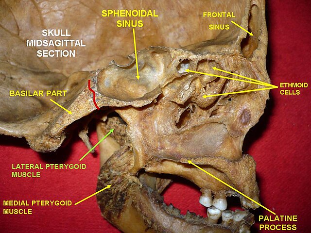
Inferior surface  -contributes to the roof of the nasal cavity and forms part of the nasal septum through articulation with the vomer.

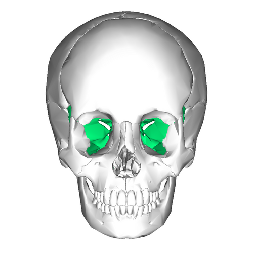
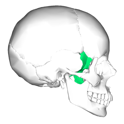
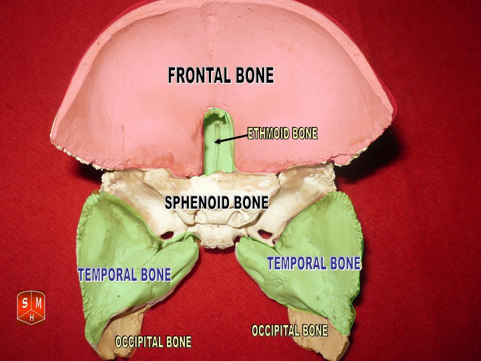
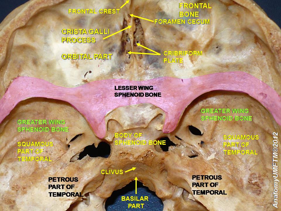
The sphenoid bone contributes surfaces to several regions:
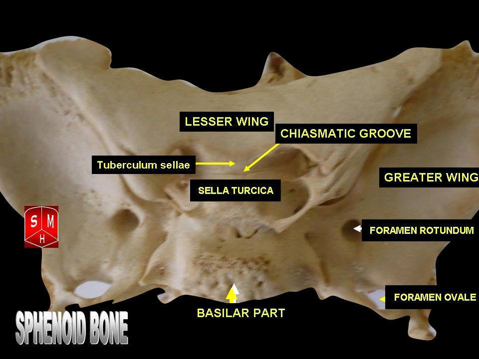
Cranial surface   forms part of the middle cranial fossa, supporting the temporal lobes of the brain.
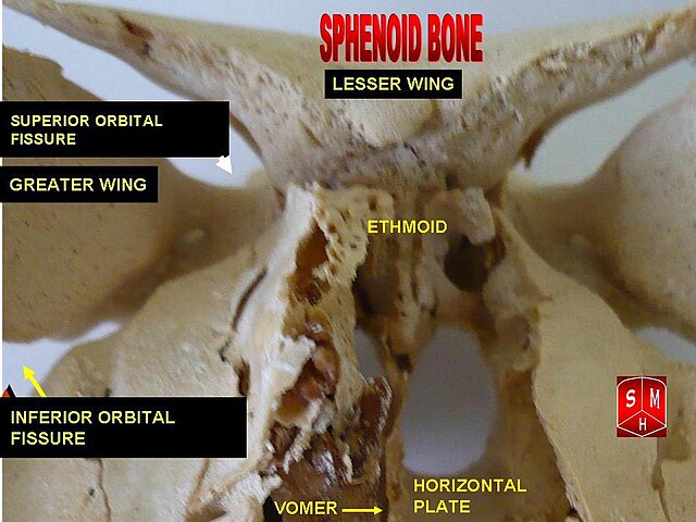
Orbital surface  contributes to the posterior wall of the orbit, supporting structures of the eye.

Temporal surface – forms part of the temporal fossa, which accommodates the temporalis muscle.

Infratemporal surface – forms part of the infratemporal fossa, a region containing important muscles and vessels associated with mastication.

Exam Question

Describe the principal surfaces of the sphenoid bone and correlate each with the cranial cavity, orbit, nasal cavity, temporal fossa, and infratemporal fossa, emphasizing how these surfaces support adjacent structures and define key anatomical boundaries.

Landmarks & Foramina

Several critical openings are located within the sphenoid bone:

Optic canal – transmits the optic nerve (CN II) and ophthalmic artery to the orbit.

Superior orbital fissure – transmits cranial nerves III, IV, V₁, and VI, as well as ophthalmic veins.

Foramen rotundum– transmits the maxillary nerve (CN V₂).

Foramen ovale– transmits the mandibular nerve (CN V₃).

Foramen spinosum– transmits the middle meningeal artery, which supplies the dura mater.

Exam Question

Examine the major landmarks and foramina of the sphenoid bone, including the sella turcica, optic canal, superior orbital fissure, and foramina rotundum, ovale, and spinosum, and explain the structural, neurovascular, and clinical importance of each.

Articulations

The sphenoid bone articulates with 12 bones, reflecting its central role within the skull.

These include:

Frontal bone; Parietal bones

Temporal bones; Occipital bone

Ethmoid bone; Zygomatic bones

Palatine bones; Vomer

Through these articulations, the sphenoid integrates the cranial base, orbit, nasal cavity, and facial skeleton.

Exam Question

The sphenoid bone articulates with numerous cranial and facial bones. Discuss these articulations in detail and explain how they establish the sphenoid as the central keystone of the skull, integrating the cranial base, orbit, nasal cavity, and facial skeleton into a unified framework.

FUNCTIONAL IMPORTANCE

Cranial Integration

The sphenoid bone serves as the central structural keystone of the cranial base, uniting the frontal, temporal, parietal, occipital, and ethmoid bones into a single mechanically integrated framework. 

Because of its central position, it contributes not only to skeletal continuity but also to the spatial organization of the anterior, middle, and posterior cranial regions. 

In this way, the sphenoid is fundamental to maintaining the architectural stability of the skull and the internal arrangement of adjacent neurovascular compartments.

Endocrine Support

The body of the sphenoid contains the sella turcica, a specialized depression that lodges the pituitary gland. This makes the sphenoid essential for structural protection of one of the most important endocrine control centers in the body. 

By enclosing and supporting the pituitary within the hypophyseal fossa, the sphenoid provides a stable osseous environment for a gland that regulates growth, metabolism, reproduction, stress response, and multiple hormonal axes

Neurovascular Transmit

The sphenoid bone contains numerous foramina, canals, and fissures that permit the orderly transmission of critical neurovascular structures between the cranial cavity, orbit, face, and infratemporal region. These include the optic canal, superior orbital fissure, and foramina rotundum, ovale, and spinosum. 

Through these openings, the sphenoid functions as a major neurovascular gateway, allowing cranial nerves and vessels to reach their targets while preserving organized compartmental anatomy at the skull base.

Orbital Formation

The lesser and greater wings of the sphenoid contribute significantly to the posterior walls of the orbit and to the floor of the middle cranial fossa. 

Through this dual role, the sphenoid participates in both orbital support and intracranial protection. It helps separate the orbital contents from the cranial cavity while also providing a rigid framework that supports visual pathways and protects delicate structures related to the eye and optic nerve.

CLINICAL RELEVANCE

Pituitary Expansion

Because the pituitary gland lies within the sella turcica, lesions such as pituitary adenomas may expand superiorly and compress the optic chiasm, producing characteristic visual disturbances, especially bitemporal hemianopia. This relationship makes the sphenoid clinically central in neuroendocrine and skull base pathology. 

Even small changes in sellar dimensions may have major consequences because of the close packing of endocrine, neural, and vascular structures in this region.

Cavernous Pathology

The sphenoid body lies in close relation to the cavernous sinus, making sphenoid-region disease highly significant for cranial nerve function. 

Lesions in this area may affect cranial nerves III, IV, V1, V2, and VI, as well as the internal carotid artery, producing ophthalmoplegia, facial sensory loss, diplopia, and potentially life-threatening vascular complications. 

This makes the sphenoid one of the most clinically sensitive bones of the cranial base.

Basal Trauma

Fractures involving the sphenoid may disrupt major skull base pathways and damage nearby structures such as the optic nerve, internal carotid artery, or cavernous sinus. Because the sphenoid forms a central part of the cranial base, traumatic injury here may produce complex neurological deficits, orbital complications, or vascular compromise. 

Sphenoid fractures are therefore clinically important not merely as bone injuries, but as markers of potentially severe intracranial damage.

Sinus Spread

The sphenoidal sinus lies within the body of the sphenoid and is closely related to the pituitary gland, optic nerve, cavernous sinus, and internal carotid artery. As a result, sphenoidal sinus infection is particularly important because inflammation or infection may extend to adjacent intracranial or orbital structures.

This explains why sphenoid sinus disease, although less common than other sinus pathology, may carry disproportionately serious neurological and ophthalmic consequences.

SUMMARY TABLE

Scroll to Top