Occipital Bone
The occipital bone is an unpaired bone forming the posterior wall and posterior portion of the cranial base of the skull. It encloses and protects the cerebellum, brainstem, and posterior parts of the cerebral hemispheres, and constitutes the major structural link between the cranial cavity and the vertebral column.
CORE


Overview
The most prominent feature of the occipital bone is the foramen magnum, a large opening through which the medulla oblongata continues as the spinal cord, along with the passage of the vertebral arteries, spinal roots of the accessory nerve (CN XI), and meninges.
Because of this relationship, the occipital bone plays a fundamental role in the craniovertebral junction, enabling articulation between the skull and the first cervical vertebra (atlas).
ANATOMY
Parts
The occipital bone is anatomically divided into 4 major parts arranged around the foramen magnum:
Squamous parts
The squamous part is the large posterior portion of the bone located behind the foramen magnum. It forms the posterior wall of the cranial vault and provides attachment sites for several muscles and ligaments of the neck.
Basilar part
The basilar part lies anterior to the foramen magnum and forms part of the cranial base.
Its superior surface slopes upward to form the clivus, a smooth area supporting the pons and medulla oblongata of the brainstem.
Anteriorly, the basilar part articulates with the body of the sphenoid bone, forming the spheno-occipital synchondrosis, an important growth region of the cranial base during development.
Lateral (Condylar part)
The lateral parts lie on each side of the foramen magnum and contain the occipital condyles, which articulate with the superior articular facets of the atlas (C1 vertebra). These articulations form the atlanto-occipital joints, which permit movements such as flexion and extension of the head (“nodding”) and slight lateral tilting. Located superior to the occipital condyles is the hypoglossal canal, which transmits the hypoglossal nerve (cranial nerve XII) responsible for motor innervation of the tongue muscles.
Exam Question
Explain the anatomical organization of the occipital bone around the foramen magnum, integrating the structural roles of the squamous, basilar, and lateral (condylar) parts, and correlate each with their contributions to cranial protection, brainstem support, and craniocervical articulation.
Surfaces
External surface
Forms the posterior aspect of the skull and provides attachment sites for numerous muscles and ligaments involved in head and neck movement.
Internal surface
Forms part of the posterior cranial fossa, accommodating the cerebellum and brainstem, and contains grooves for major dural venous sinuses.
Exam Question
Analyze the external and internal surfaces of the occipital bone, correlating external muscular attachment sites with head posture and movement, and internal features with the organization of the posterior cranial fossa and dural venous sinus pathways.
Borders
The occipital bone possesses several borders that articulate with neighboring cranial bones.
Lambdoid Border – superior border articulates with the parietal bones, forming the lambdoid suture.
Mastoid Border – lateral borders articulate with the temporal bones, contributing to the occipitomastoid sutures.
Basilar Border – anterior border articulates with the sphenoid bone, forming part of the cranial base.
Exam Question
Discuss the borders of the occipital bone in relation to major cranial sutures, emphasizing their role in skull stability, force transmission across the posterior cranial vault, and developmental integration with adjacent bones
Articulations
The occipital bone articulates with 4 bones:
Parietal bones
Temporal bones
Sphenoid bone
Atlas (C1 vertebra)
Through these articulations, the occipital bone connects the cranial vault with the vertebral column,forming the structural basis of the craniovertebral junction.
Exam Question
Evaluate the articulations of the occipital bone, particularly the occipital condyles, and explain how these joints facilitate controlled head movement while transmitting axial loads between the skull and vertebral column.
Landmarks
Important anatomical landmarks include:
Foramen magnum – passage for the brainstem and vertebral arteries
Occipital condyles – articulate with the atlas
External occipital protuberance – attachment for ligamentum nuchae
Superior and inferior nuchal lines – muscle attachment sites
Internal occipital protuberance – intersection of venous sinus grooves
Hypoglossal canal – passage for hypoglossal nerve (CN XII)
Exam Question
Describe the key anatomical landmarks of the occipital bone, including the foramen magnum, nuchal lines, and hypoglossal canal, and correlate their structural features with neurovascular transmission, muscle attachment, and clinical relevance.
FUNCTIONAL IMPORTANCE
Posterior Cranial Protection
The occipital bone forms the posterior part of the cranial vault and the posterior cranial fossa, providing rigid protection for the cerebellum, brainstem, and occipital lobes.
Through its curved architecture and dense cortical framework, it resists external forces while safeguarding structures essential for coordination, autonomic regulation, and visual processing.
Craniovertebral Continuity
Through the foramen magnum, the occipital bone establishes the critical anatomical transition between the cranial cavity and vertebral canal.
This aperture permits continuity of the medulla oblongata with the spinal cord, as well as transmission of vertebral arteries, meninges, and associated neurovascular structures, thereby preserving structural and functional integrity of the central neuroaxis.
Atlanto-Occipital Articulation
The occipital condyles articulate with the superior articular facets of the atlas to form the atlanto-occipital joints, which permit controlled flexion and extension of the head while maintaining craniovertebral stability.
This articulation is biomechanically essential for balanced head support, transmission of axial loads to the cervical spine, and precise postural adjustment.
Musculoskeletal Postural Integration
The external surface of the occipital bone provides extensive attachment sites for nuchal and suboccipital musculature, including muscles responsible for head extension, rotation, and stabilization.
Through these attachments, the bone serves as a major anchor for postural mechanics, integrating cranial stability with coordinated cervical movement and maintenance of upright head position.
CLINICAL RELEVANCE
Foramen Magnum Compression
Pathology involving the foramen magnum, including congenital anomalies, mass lesions, or downward herniation, may compress the medulla oblongata and upper cervical spinal cord, producing severe neurological compromise.
Because this region contains vital autonomic and long-tract structures, compression may lead to respiratory failure, cardiovascular instability, lower cranial nerve dysfunction, and motor deficits.
Occipital Condyle fracture
Fractures of the occipital condyles, typically associated with high-energy trauma, may destabilize the craniovertebral junction and threaten the integrity of adjacent neural and ligamentous structures.
Such injuries are clinically significant because they may compromise alignment between skull and cervical spine, with risk of upper spinal cord injury and neurovascular disturbance.
Hypoglossal Canal Involvement
Lesions involving the hypoglossal canal may affect the hypoglossal nerve, producing ipsilateral weakness of tongue musculature.
Clinically, this may present with tongue deviation on protrusion, dysarthria, and impaired swallowing, making the occipital bone highly relevant in skull base pathology and lower cranial nerve syndromes.
Neurosurgical Significance
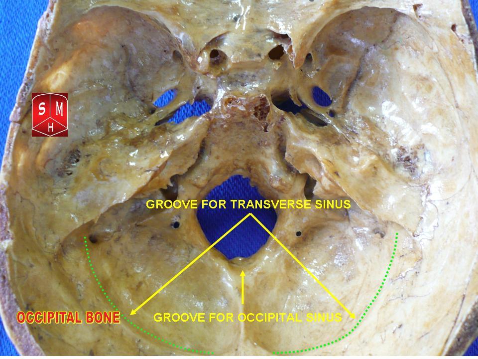
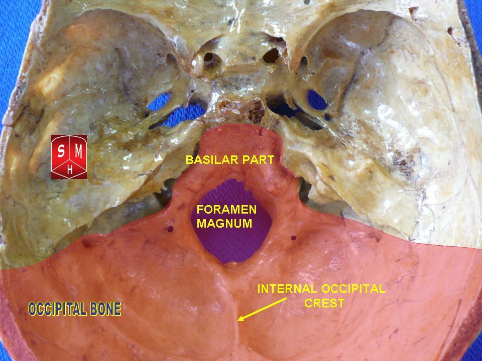
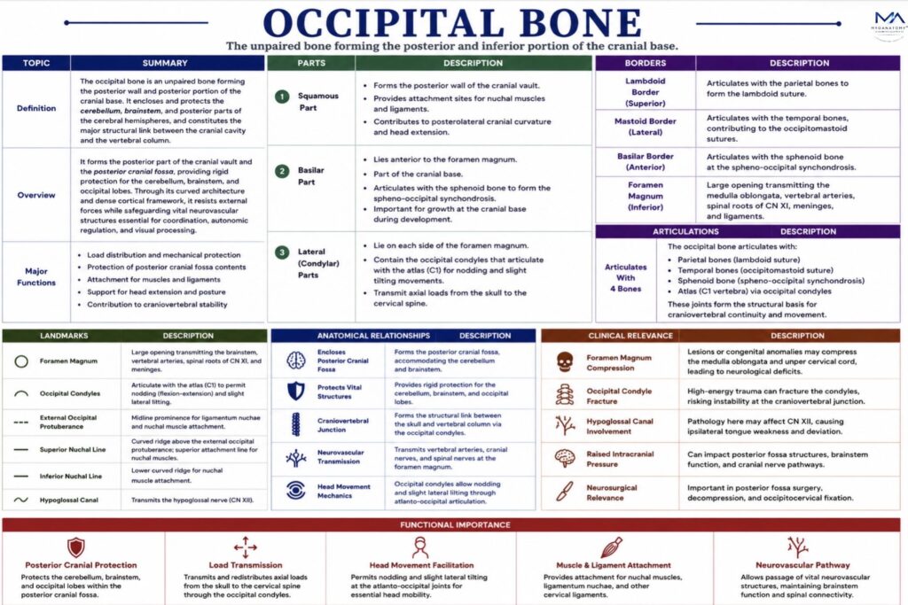
Because the occipital bone encloses the posterior cranial fossa and lies in close relation to dural venous sinuses, cerebellar structures, and the craniovertebral junction, pathology or surgical intervention in this region carries substantial risk.
Fracture, mass effect, or operative error may result in venous injury, raised intracranial pressure, cerebellar dysfunction, or brainstem compression, making precise anatomical knowledge essential in trauma care and neurosurgery
SUMMARY TABLE
