Frontal Bone
The frontal bone is a large, unpaired bone forming the anterior portion of the cranial vault, the roof of the orbits, and a substantial part of the anterior cranial base. It contributes significantly to both the neurocranium and the viscerocranium, linking the cranial cavity with the facial skeleton.
CORE


Overview
Superiorly, the frontal bone forms the forehead and anterior cranial vault, protecting the frontal lobes of the brain, which are responsible for higher cognitive functions, voluntary motor control, and behavioral regulation.
Inferiorly, it participates in the formation of the orbital cavities, separating the orbital contents from the anterior cranial fossa.
Through its articulations with numerous cranial and facial bones, the frontal bone serves as an important structural element of the upper facial skeleton and cranial base.
ANATOMY
Parts
The frontal bone is anatomically divided into 3 major parts:
Squamous part; Orbital part; Nasal part
These regions correspond to the bone’s contributions to the cranial vault, orbital cavity, and nasal region.
Squamous Part
The squamous part forms the vertical anterior portion of the skull, constituting the forehead and the anterior wall of the cranial vault. It protects the frontal lobes of the cerebral hemispheres and provides attachment sites for muscles of the scalp and facial expression.
Orbital Part
The orbital part consists of two horizontal plates known as the orbital plates, which form the roof of the orbits and the floor of the anterior cranial fossa. These plates separate the orbital cavities from the cranial cavity and provide structural protection for the eyes and associated neurovascular structures.
Nasal Part
The nasal part lies between the orbital plates and contributes to the root of the nose and superior portion of the nasal cavity. It articulates with the nasal bones and maxillae, helping form the upper boundary of the nasal skeleton.
Exam Question
How does the anatomical division of the frontal bone into squamous, orbital, and nasal parts reflect its structural integration within the cranial vault, orbital cavity, and nasal region, and how does each part contribute to the organization of the anterior skull?
Surfaces
The frontal bone presents 2 principal surfaces: an external surface and an internal surface.
External Surface
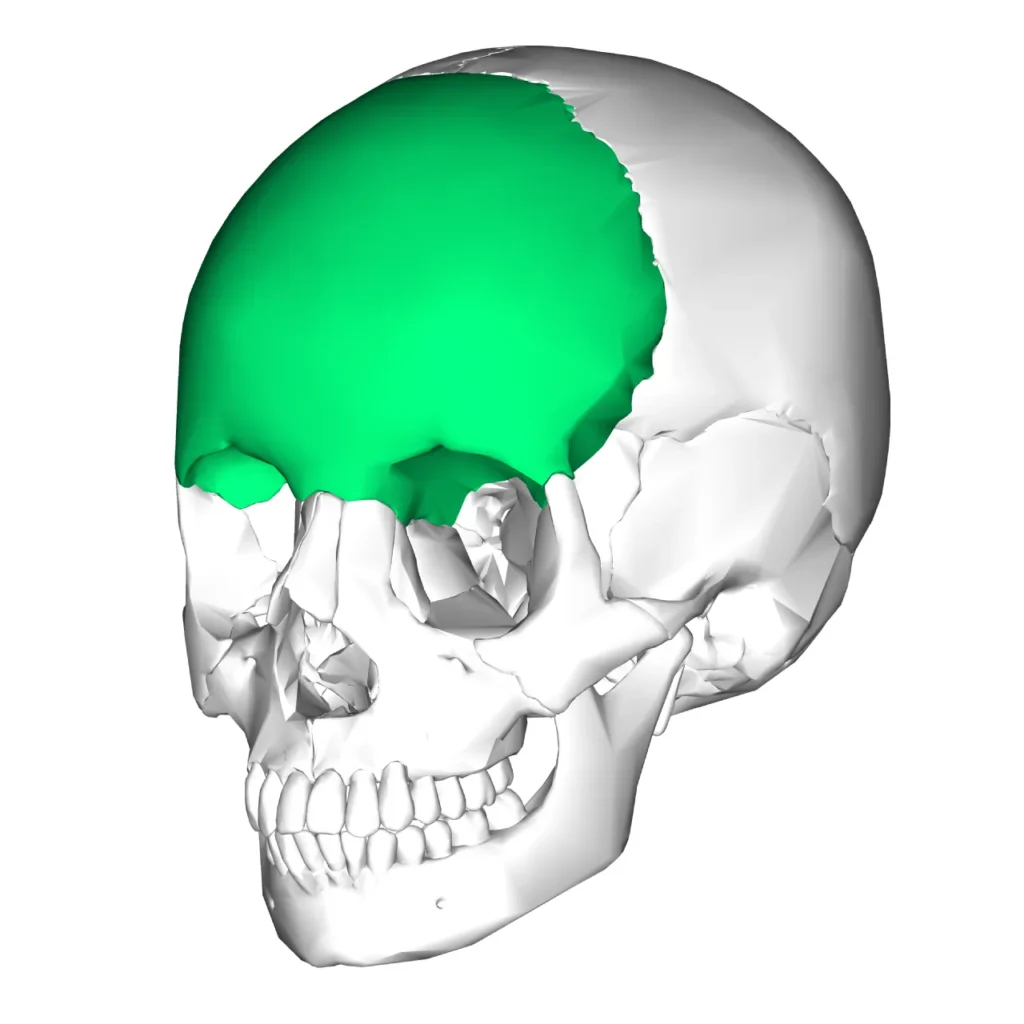
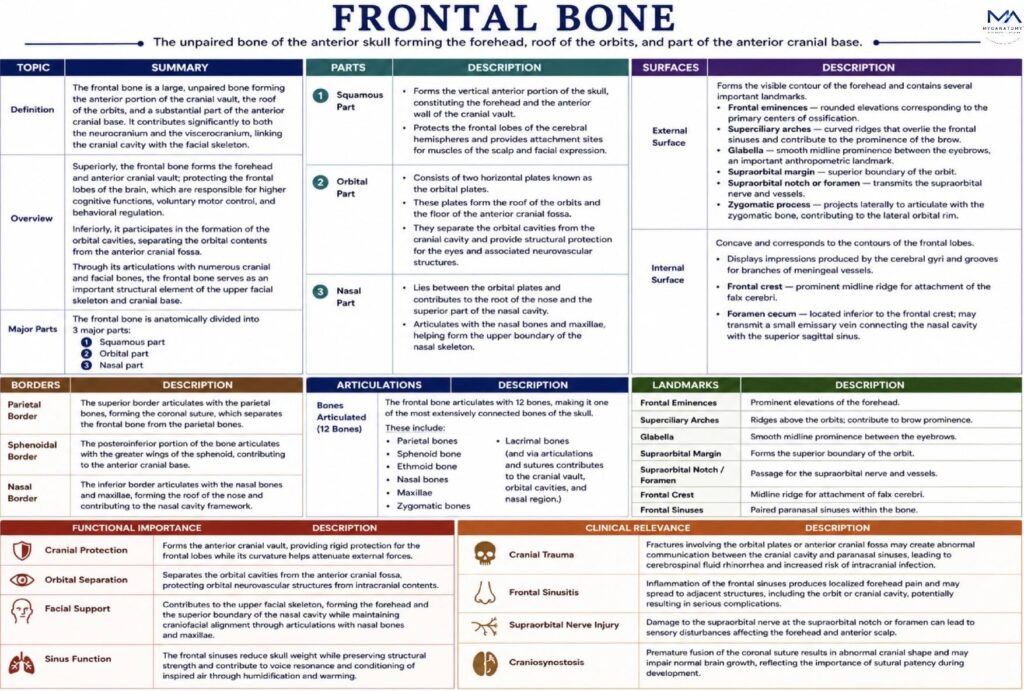
The external surface forms the visible contour of the forehead and contains several prominent landmarks.
The frontal eminences represent rounded elevations corresponding to the primary centers of ossification of the bone.
Below these lie the superciliary arches, curved ridges that overlie the frontal sinuses and contribute to the prominence of the brow.
Between the superciliary arches lies the glabella, a smooth midline prominence that serves as an important anthropometric landmark.
Inferiorly, the supraorbital margin forms the superior boundary of the orbit. Along this margin lies the supraorbital notch or foramen, which transmits the supraorbital nerve and vessels, supplying the forehead and anterior scalp.
The zygomatic process of the frontal bone projects laterally to articulate with the zygomatic bone, contributing to the lateral orbital rim.
Internal Surface
The internal surface of the frontal bone is concave and corresponds to the contours of the frontal lobes of the brain.
This surface displays impressions produced by the cerebral gyri and grooves for branches of the meningeal vessels.
A prominent midline ridge known as the frontal crest provides attachment for the falx cerebri, a dural fold separating the cerebral hemispheres.
Inferior to the frontal crest lies the foramen cecum, which occasionally transmits a small emissary vein connecting the nasal cavity with the superior sagittal sinus.
Exam Question
How do the internal surface features of the frontal bone reflect its anatomical relationship with the cerebral hemispheres and meningeal vessels, and how do these impressions organize neurovascular pathways within the cranial cavity?
Borders
The frontal bone possesses several borders that participate in major cranial sutures.
Parietal Border
The superior border articulates with the parietal bones, forming the coronal suture, which separates the frontal bone from the parietal bones.
Sphenoidal Border
The posteroinferior portion of the bone articulates with the greater wings of the sphenoid, contributing to the anterior cranial base.
Nasal Border
The inferior border articulates with the nasal bones and maxillae, forming the roof of the nose and contributing to the nasal cavity framework.
Exam Question
How do the structural orientation and articulating edges of the frontal bone borders contribute to the formation of major cranial sutures, and how do these relationships influence cranial alignment and growth?
Articulations
The frontal bone articulates with 12 bones, making it one of the most extensively connected bones of the skull.
These include:
Parietal bones
Sphenoid bone
Ethmoid bone
Nasal bones
Maxillae
Zygomatic bones
Lacrimal bones
Through these articulations, the frontal bone contributes to the formation of the cranial vault, orbital cavities, and nasal region.
Exam Question
How do the articulations of the frontal bone with adjacent cranial and facial bones establish continuity within the cranial vault, and how do these connections contribute to overall structural integration of the skull?
Landmarks
Important anatomical landmarks associated with the frontal bone include:
Frontal eminences – prominent elevations of the forehead
Superciliary arches – ridges above the orbits
Glabella – smooth midline prominence between the eyebrows
Supraorbital margin – superior boundary of the orbit
Supraorbital notch or foramen – passage for supraorbital nerve and vessels
Frontal crest attachment site for the falx cerebri
Frontal sinuses – paranasal sinuses located within the bonE. The frontal sinuses vary in size and contribute to reduction of skull weight, resonance of the voice, and conditioning of inspired air.
Exam Question
How do the major anatomical landmarks of the frontal bone correspond to muscle attachments, neurovascular transmission pathways, and key reference points for anatomical orientation?
FUNCTIONAL IMPORTANCE
Cranial Protection
The frontal bone forms the anterior cranial vault, providing rigid protection for the frontal lobes, which are responsible for executive function, voluntary motor activity, and behavioral regulation, while its curvature helps attenuate external forces.
Orbital Seperation
The frontal bone forms the anterior cranial vault, providing rigid protection for the frontal lobes, which are responsible for executive function, voluntary motor activity, and behavioral regulation, while its curvature helps attenuate external forces.
Facial Support
The frontal bone contributes to the upper facial skeleton, forming the forehead and the superior boundary of the nasal cavity, thereby maintaining midfacial stability and craniofacial alignment through its articulations with the nasal bones and maxillae.
Sinus Function
The frontal sinuses reduce skull weight while preserving structural strength and contribute to resonance of the voice and conditioning of inspired air through humidification and warming.
CLINICAL RELEVANCE
Cranial Trauma
Fractures involving the orbital plates or anterior cranial fossa may create abnormal communication between the cranial cavity and paranasal sinuses, leading to cerebrospinal fluid rhinorrhea and increased risk of intracranial infection.
Frontal Sinusitis
Inflammation of the frontal sinuses produces localized forehead pain and may spread to adjacent structures, including the orbit or cranial cavity, potentially resulting in serious complications.
Supraorbital Nerve Injury
Damage to the supraorbital nerve at the supraorbital notch or foramen can lead to sensory disturbances affecting the forehead and anterior scalp.
Craniosynostosis
Premature fusion of the coronal suture results in abnormal cranial shape and may impair normal brain growth, reflecting the importance of sutural patency during development
SUMMARY TABLE
