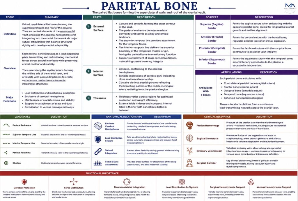
Parietal Bone
Paired, quadrilateral flat bones forming the superolateral walls and roof of the calvaria, the parietal bones are central elements of the neurocranial vault, enclosing the cerebral hemispheres and integrating into the cranial framework via major sutural articulations that balance structural rigidity with developmental adaptability.
CORE


Overview
Each parietal bone functions as a load-dispersing plate, transmitting and redistributing mechanical forces across sutural interfaces while preserving cranial contour and stability.
They meet along the sagittal suture, forming the midline axis of the cranial vault, and articulate with surrounding bones to create a continuous protective enclosure for intracranial structures
ANATOMY
Surfaces
External Surface
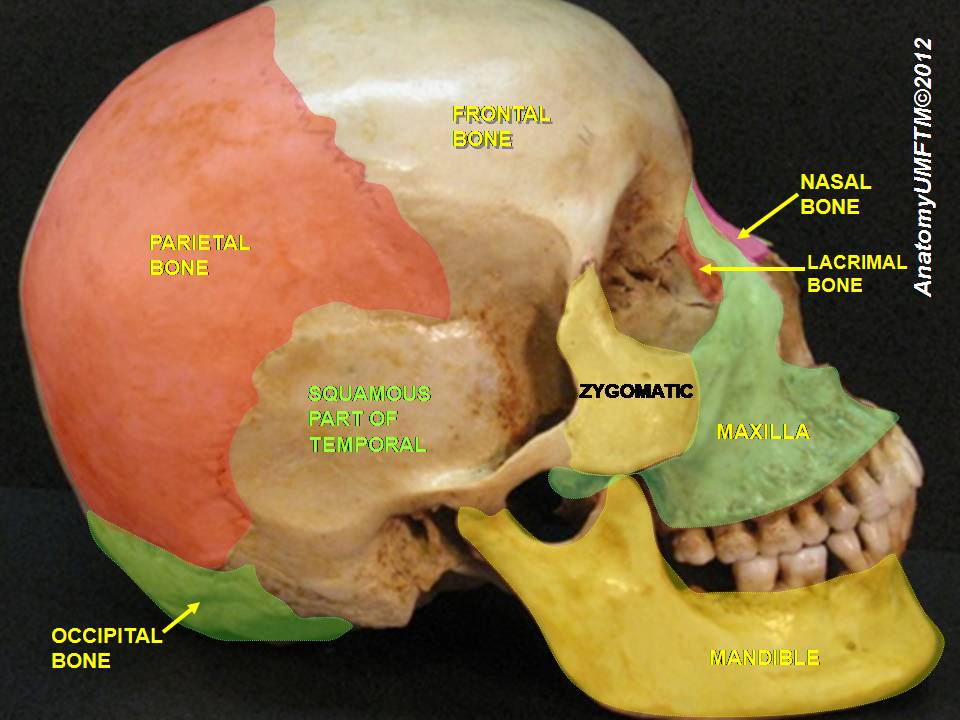
Convex and smooth, forming the outer contour of the skull. The parietal eminence denotes maximal convexity and serves as a key anatomical landmark.
The superior temporal line provides attachment for the temporal fascia, while the inferior temporal line defines the superior boundary of the temporalis muscle origin, linking the parietal bone to masticatory function.
This surface also supports attachment of scalp connective tissues, maintaining structural continuity of the cranial covering.
Internal Surface
Concave, conforming to the cerebral hemispheres, with impressions of cerebral gyri indicating close anatomical relationship.
Distinct arterial grooves reflect the branching pattern of the middle meningeal artery, radiating from the pterional region.
Shallow depressions near the sagittal region correspond to arachnoid granulations, representing sites of cerebrospinal fluid resorption into the superior sagittal sinus.
Exam Question
How do the internal surface features of the parietal bone reflect its relationship with intracranial structures, and what is the functional significance of these impressions in neurovascular organization.
Borders
Superior (Sagittal) Border – forms the sagittal suture, permitting longitudinal cranial growth and maintaining midline alignment
Anterior (Frontal) Border – forms the coronal suture, regulating anterior–posterior cranial expansion
Posterior (Occipital) Border – forms the lambdoid suture, contributing to posterior vault integrity
Inferior (Squamous) Border – forms the squamous suture with the temporal bone; anteroinferiorly contributes to the pterion, a structurally thin junction
Exam Question
Analyze how the orientation and articulation of the parietal bone borders contribute to cranial growth patterns and mechanical force distribution across the calvarial sutures.
Articulations
Each parietal bone articulates with five bones:
Contralateral parietal bone – sagittal suture
Frontal bone – coronal suture
Occipital bone – Lambdoid suture
Temporal bone – squamous suture
Sphenoid bone – pterional region
These articulations form a continuous load-transmitting network across the cranial vault.
Exam Question
Explain how the articulations of the parietal bone form an integrated biomechanical network within the cranial vault, and discuss their role in maintaining structural integrity under dynamic loading conditions.
Landmarks
Parietal eminence – maximal convexity
Superior temporal line – temporal fascia attachment
Inferior temporal line – superior limit of temporalis origin
Parietal foramina – emissary veins → superior sagittal sinus
Obelion – midline landmark between parietal foramina
Exam Question
Correlate the major anatomical landmarks of the parietal bone with their functional and clinical significance, particularly in relation to muscle attachment, venous drainage, and neurosurgical orientation.
FUNCTIONAL IMPORTANCE
Cerebral Protection
The parietal bones form a major portion of the calvaria, acting as rigid protective plates shielding the cerebral hemispheres. Their thickness and curvature reduce transmission of external forces, preserving cortical regions responsible for sensory integration and spatial processing.
Force Distribution
The convex architecture functions as a biomechanical load-dispersing system, converting focal impacts into distributed tensile and compressive stresses across sutural interfaces. The diploic structure enables energy absorption and stress attenuation, minimizing focal intracranial injury.
Musculoskeletal Integration
The superior and inferior temporal lines transmit forces from the temporalis muscle and temporal fascia, integrating cranial bone with masticatory biomechanics. This allows efficient force transfer during jaw elevation without compromising cranial structural stability.
Venous Hemodynamic
Parietal foramina transmit valveless emissary veins, enabling bidirectional flow between extracranial veins and the superior sagittal sinus.
This supports intracranial venous pressure regulation and maintains cerebral venous outflow equilibrium.
CLINICAL RELEVANCE
Pteron Hemorrhage
The pterion overlies the anterior branch of the middle meningeal artery. Fracture leads to arterial rupture and rapid epidural hematoma formation, causing acute intracranial pressure elevation and risk of transtentorial herniation
Sagital Synostosis
Premature fusion of the sagittal suture abolishes transverse growth, producing scaphocephaly. This alters cranial geometry and may affect intracranial volume adaptation and neurodevelopment.
Emissary Vein Spread
Valveless emissary veins create a pathway for retrograde infection spread from scalp to intracranial venous sinuses, predisposing to venous sinus thrombosis or intracranial infection.
Surgical Corridor
The parietal bone is a key site for craniotomy, but its internal surface contains grooves of the middle meningeal vessels and proximity to the superior sagittal sinus. Surgical inaccuracy risks vascular injury and dural compromise.
SUMMARY TABLE
