Bone Ossification
MYO CORE
Osteogenesis Basis
Bone ossification is a tightly regulated process by which mesenchymal tissue is transformed into mineralized bone through coordinated cellular differentiation, osteoid synthesis, and hydroxyapatite deposition, producing a structurally competent and metabolically active skeletal framework.
OVERVIEW
It underlies embryonic development, postnatal growth, fracture repair, and lifelong skeletal maintenance, ensuring mechanical stability, protection, and calcium-phosphate homeostasis.
This process is mediated by integrated activity of osteogenic lineage cells within a vascularized environment, where osteoblasts deposit a collagen-rich matrix that subsequently mineralizes, conferring rigidity and load-bearing capacity, while osteocytes coordinate adaptive responses and osteoclasts enable structural renewal.
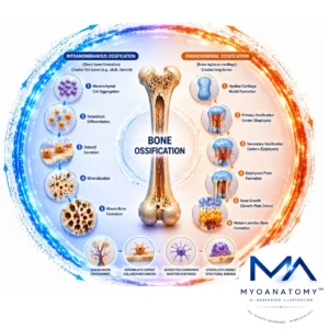
Ossification occurs via intramembranous and endochondral pathways, which differ in developmental sequence but converge in the formation of lamellar bone optimized for mechanical function. Throughout life, ossification remains dynamically regulated by mechanical forces, hormonal control, and molecular signaling, ensuring continuous adaptation, structural integrity, and metabolic balance of bone tissue.
ANATOMY
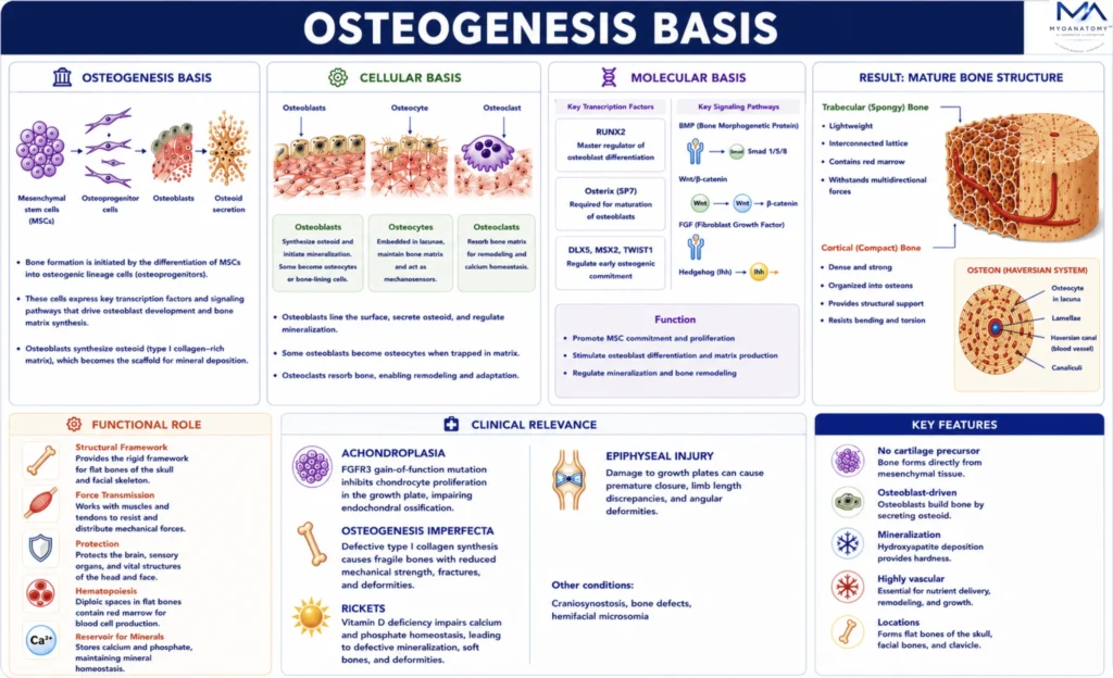
Osteogenesis Basis
Bone formation is initiated by the differentiation of mesenchymal stem cells into osteogenic lineage cells. This differentiation is governed by several key transcription factors and signaling pathways that regulate osteoblast development and bone matrix synthesis.
Cellular Basis
Osteogenesis is initiated by the differentiation of mesenchymal stem cells (MSCs) into osteogenic lineage cells under tightly regulated developmental programs. These cells sequentially give rise to osteoprogenitors, osteoblasts, osteocytes, and osteoclasts, forming a coordinated cellular network responsible for bone formation and remodeling.
Osteoblasts synthesize osteoid and initiate mineralization, while embedded osteocytes function as mechanosensors, regulating bone turnover in response to mechanical and metabolic stimuli. Osteoclasts, derived from the hematopoietic lineage,mediate bone resorption, enabling continuous structural adaptation.
The dynamic balance between osteoblast-mediated formation and osteoclast-mediated resorption ensures skeletal integrity, functional adaptation, and maintenance of bone mass.
Exam Question
Explain the coordinated roles of osteoblasts, osteocytes, and osteoclasts in maintaining bone homeostasis, and describe how disruption of their balance leads to pathological bone remodeling.
Molecular Basis
Osteogenesis is regulated by tightly integrated transcription factors and signaling pathways that control osteogenic commitment, differentiation, and matrix production.
The transcription factor RUNX2is essential for early osteoblast differentiation, while Osterix (SP7) drives osteoblast maturation. BMP signaling promotes osteogenic lineage commitment, and the Wnt/β-catenin pathway enhances osteoblast proliferation and bone formation.
Indian hedgehog (Ihh) regulates chondrocyte activity and growth plate organization during endochondral ossification, while fibroblast growth factors (FGFs) contribute to skeletal patterning and longitudinal growth.
These pathways interact to precisely regulate gene expression, cellular differentiation, and matrix synthesis, ensuring coordinated bone formation and development.
Exam Question
Discuss the roles of RUNX2, Osterix, BMP, and Wnt/β-catenin signaling in osteoblast differentiation, and explain how their interaction coordinates bone formation during development and growth.
FUNCTIONAL ROLE
Structural Framework
Ossification establishes the fundamental architectural and mechanical basis of the skeleton by transforming compliant connective tissues into rigid, mineralized bone.
This process creates the load-bearing framework required for posture, stability, and coordinated movement, while organizing cortical and trabecular bone into an optimized structure that balances strength, weight, and mechanical efficiency.
Force Transmission
Ossified bone functions as an effective lever system for skeletal muscles.
The rigidity achieved through mineralization enables efficient transmission of muscular forces via tendons across joints, allowing controlled movement. Simultaneously, the formation of reinforced entheses ensures safe transfer of tensile forces from muscles and ligaments to bone without structural failure.
Joint formation
Ossification ensures the precise development of epiphyses and articular surfaces, which is essential for the formation of stable, congruent synovial joints.
Proper shaping of these surfaces allows efficient load distribution, minimizes friction during movement, and preserves long-term joint function and mechanical integrity.
Growth Adaption
Through growth plate activity, ossification enables longitudinal bone growth and proportional development of the body.
Additionally, it provides the structural substrate for lifelong bone remodeling, allowing continuous adaptation to mechanical loading, maintenance of skeletal strength, and preservation of functional performance under changing physiological demands.
CLINICAL RELEVANCE
Achondroplasia
Achondroplasia is the most common form of disproportionate dwarfism and results from a mutation in the fibroblast growth factor receptor-3 (FGFR3) gene. This mutation inhibits chondrocyte proliferation within the epiphyseal growth plate, thereby impairing endochondral ossification.
As a result, longitudinal bone growth is reduced, producing shortened limbs while the axial skeleton remains relatively preserved.
Osteogenesis Imperfecta
Osteogenesis imperfecta is a genetic disorder characterized by defective synthesis of type I collagen, the principal structural protein of bone matrix.
Because collagen provides the scaffold for mineral deposition during ossification, defective collagen formation leads to fragile bones with reduced mechanical strength. Clinically, this condition presents with recurrent fractures, skeletal deformities, and decreased bone density.
Rickets
Rickets is a disorder of defective bone mineralization, most commonly caused by vitamin D deficiency. Insufficient vitamin D impairs calcium and phosphate homeostasis, preventing proper mineral deposition within the osteoid matrix produced during ossification.
The resulting bones are soft and structurally weak, leading to skeletal deformities such as bowing of the long bones and widening of the growth plates.
Epiphyseal Injury
Trauma to the epiphyseal (growth) plates during childhood can disrupt the normal process of endochondral ossification responsible for longitudinal bone growth. Damage to the growth plate may lead to premature closure, asymmetric growth, or permanent limb length discrepancies.
From a clinical perspective, understanding the mechanisms of ossification is essential in multiple medical disciplines, including orthopedics, pediatrics, endocrinology, and rehabilitation medicine. Accurate knowledge of these processes allows clinicians to diagnose developmental skeletal disorders, evaluate and develop therapeutic strategies.
SUMMARY TABLE