Peripheral Nerves
MYO CORE
Nerve Classification
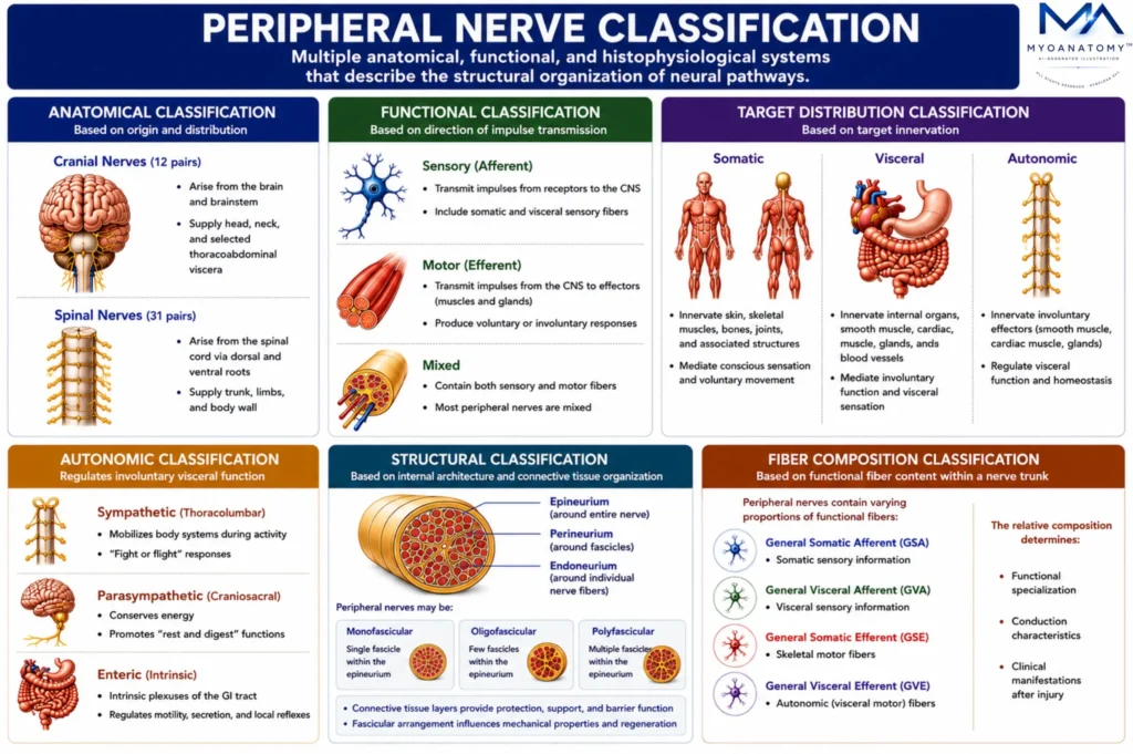
Peripheral nerves are classified according to their functional organization, anatomical origin, target innervation, autonomic components, and fiber composition, reflecting the hierarchical structural and physiological specialization of the peripheral nervous system.
OVERVIEW
Peripheral nerve classification encompasses multiple anatomical, functional, and histophysiological systems that describe the structural organization of neural pathways.
Functionally, nerves are classified as sensory (afferent), motor (efferent), or mixed; anatomically as cranial or spinal nerves; and by target distribution into somatic and visceral divisions.
Autonomic fibers are further classified into sympathetic, parasympathetic, and enteric components that regulate involuntary visceral function.
At the microscopic level, peripheral nerves are further classified according to fiber modality, axonal diameter, degree of myelination, and conduction velocity, distinguishing specialized fiber populations optimized for proprioception, somatic sensation, motor control, autonomic regulation, and nociception.
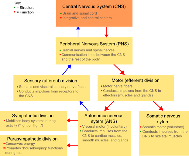
“Diagram showing the divisions of the nervous system” by Fuzzform, via Wikimedia Commons. Licensed under CC BY-SA 3.0.
Exam Question
Evaluate how anatomical, functional, autonomic, and histophysiological classifications collectively determine the structural organization, conduction properties, and specialized physiological roles of peripheral nerves.
ANATOMY
Anatomical Classification
Peripheral nerves are anatomically classified according to their origin, regional distribution, and segmental organization.
Cranial nerves arise from the brain and brainstem, supplying the head, neck, and selected thoracoabdominal viscera, whereas
Spinal nerves originate from the spinal cord through the union of dorsal sensory and ventral motor roots before dividing into dorsal and ventral rami, communicating rami, and terminal branches.
Peripheral nerves may further remain segmental or contribute to neural plexuses, where fibers are redistributed into terminal nerves supplying the limbs.
This organization establishes dermatomal and myotomal relationships while providing the anatomical basis for localization of peripheral nerve and spinal root lesions.
Exam Question
How does the anatomical classification of peripheral nerves establish segmental innervation, plexus formation, and clinical localization of peripheral nerve lesions?
Functional Classifcation
Peripheral nerves are functionally classified according to the direction and physiological role of impulse transmission.
Afferent (sensory) nerves convey somatic and visceral information from peripheral receptors to the central nervous system, whereas
Efferent (motor) nerves transmit commands from the central nervous system to skeletal muscle, smooth muscle, cardiac muscle, and glands.
Most peripheral nerves are mixed nerves, containing integrated sensory, motor, and autonomic fibers within a common nerve trunk.
This functional organization enables simultaneous sensory perception, voluntary motor control, autonomic regulation, and reflex integration while preserving efficient bidirectional communication between peripheral tissues and the central nervous system.
Exam Question
How does the functional classification of peripheral nerves integrate afferent, efferent, and mixed neural pathways to coordinate sensory, motor, autonomic, and reflex functions?
Target Distribution Classifcation
Peripheral nerves are classified according to their target innervation into somatic and visceral systems.
Somatic nerves supply the body wall and limbs, innervating skin, skeletal muscles, bones, joints, and connective tissues responsible for conscious sensation and voluntary movement.
Visceral nerves innervate smooth muscle, cardiac muscle, glands, blood vessels, and internal organs, mediating involuntary physiological regulation while transmitting visceral sensory information essential for homeostasis.
This classification reflects embryological development, receptor specialization, and the functional distinction between voluntary somatic control and autonomic visceral regulation.
Exam Question
How does the somatic and visceral classification of peripheral nerves reflect differences in target innervation, physiological function, and neural organization?
Autonomic Classification
The autonomic component of peripheral nerves is classified into sympathetic, parasympathetic, and enteric divisions, each possessing distinct central origins, peripheral ganglia, neurotransmitters, and patterns of target innervation.
Preganglionic and postganglionic neurons form two-neuron pathways that regulate cardiovascular function, glandular secretion, smooth muscle activity, thermoregulation, and visceral homeostasis.
Although functionally distinct, these autonomic divisions operate as an integrated neuroregulatory network that continuously adapts organ function to changing physiological demands while maintaining internal homeostasis.
Exam Question
How do the structural organization and functional specializations of the sympathetic, parasympathetic, and enteric divisions coordinate autonomic regulation and visceral homeostasis?
Structural Classifcation
Peripheral nerves are structurally classified according to their internal fascicular architecture and connective tissue organization.
Individual axons are enclosed by the endoneurium, grouped into fascicles by the perineurium, and surrounded collectively by the epineurium, forming a hierarchical framework that provides mechanical protection, electrical insulation, vascular support, and selective molecular permeability.
Peripheral nerves may exhibit monofascicular, oligofascicular, or polyfascicular organization depending on their anatomical location and functional demands.
Variations in fascicular arrangement, connective tissue content, and vascular distribution influence nerve elasticity, resistance to compression, microsurgical repair, and regenerative capacity.
Exam Question
How does the structural classification of peripheral nerves optimize mechanical protection, fascicular organization, vascular support, and regenerative potential?
Fiber Composition Classifcation
Peripheral nerve fibers are classified according to functional modality, axonal diameter, degree of myelination, and conduction velocity, characteristics that collectively determine the speed, precision, and physiological specialization of neural transmission.
Functionally, fibers are categorized as general somatic afferent (GSA), general visceral afferent (GVA), general somatic efferent (GSE), and general visceral efferent (GVE), whereas the Erlanger–Gasser classification distinguishes A, B, and C fibers, with A fibers further subdivided into Aα, Aβ, Aγ, and Aδ according to progressively decreasing diameter and conduction velocity.
Large, heavily myelinated Aα fibers primarily conduct GSE motor output and GSA proprioceptive input; Aβ fiber stransmit GSA discriminative touch, vibration, and pressure; Aγ fibers provide GSE innervation to intrafusal muscle fibers, regulating muscle spindle sensitivity;
Aδ fibers carry GSA fast pain and cold sensation; B fibers represent lightly myelinated GVE preganglionic autonomic fibers; where as unmyelinated C fibers transmit GSA slow pain and warmth, GVA visceral afferent information, and GVE postganglionic autonomic signals.
Exam Question
How do functional fiber modalities (GSA, GVA, GSE, GVE) and the Erlanger–Gasser classification collectively determine the conduction properties, physiological specialization, and functional integration of peripheral nerve fibers?
Segmental & Neural Classifcation
Peripheral nerves exhibit either segmental organization or plexiform organization.
Thoracic spinal nerves largely retain their original segmental distribution, whereas the
Ventral rami of cervical, brachial, lumbar, sacral, and coccygeal regions interconnect to form neural plexuses. Within these plexuses, nerve fibers are redistributed into terminal peripheral nerves containing axons derived from multiple spinal cord segments.
This reorganization enhances functional redundancy, coordinates complex limb innervation, and minimizes complete loss of function following injury to a single spinal nerve root.
Exam Question
How does plexus formation modify segmental spinal nerve organization to optimize peripheral innervation and preserve functional redundancy following nerve injury?
SUMMARY TABLE