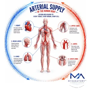
Arterial Supply
MYO CORE
Anastomosis
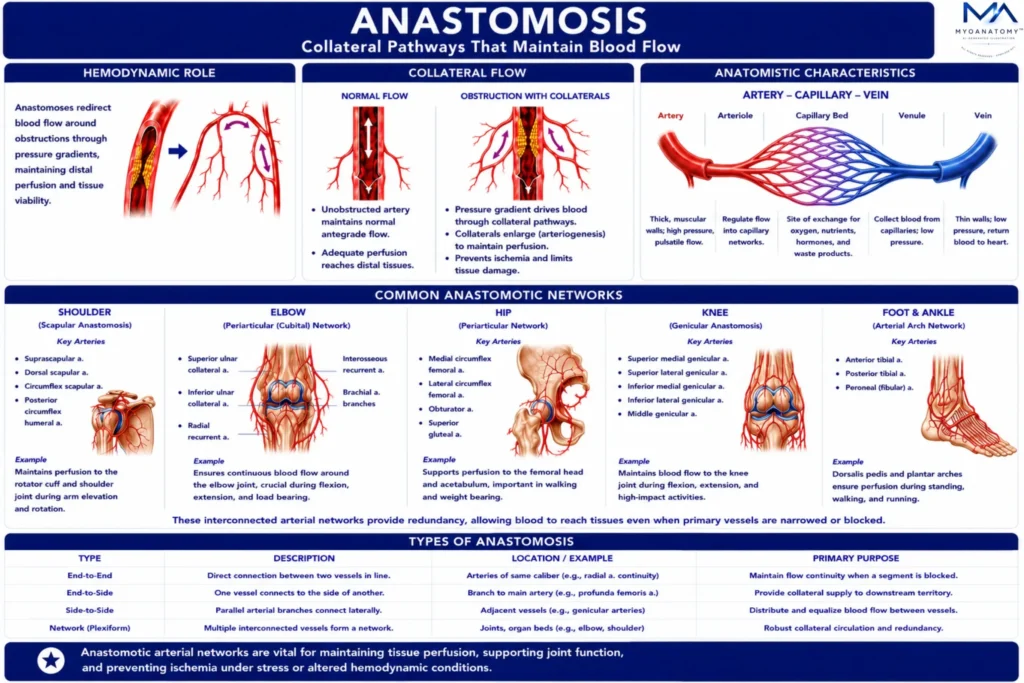
Arterial anastomoses are direct vascular connections between two or more arteries supplying the same anatomical region, forming alternative pathways for blood flow. These networks function as collateral channels that preserve tissue perfusion when the primary arterial route is narrowed, compressed, or occluded.
OVERVIEW
Hemodynamically, anastomoses enable redistribution of blood flow by allowing pressure gradients to redirect circulation through secondary pathways. In conditions of gradual arterial obstruction, these vessels can undergo adaptive enlargement (arteriogenesis), increasing their capacity to maintain distal perfusion.
This collateral circulation is critical in preventing ischemia, particularly in regions exposed to mechanical stress or variable blood supply.
Within the musculoskeletal system, arterial anastomoses are highly developed around mobile joints, where movement may transiently compromise primary vessels. These networks ensure continuous perfusion during changes in joint position and mechanical loading.
Functionally significant anastomotic networks are found around the shoulder (scapular anastomosis), elbow (periarticular arterial network), hip, and knee (genicular anastomosis), where interconnected arterial branches maintain vascular continuity and protect against ischemic injury.
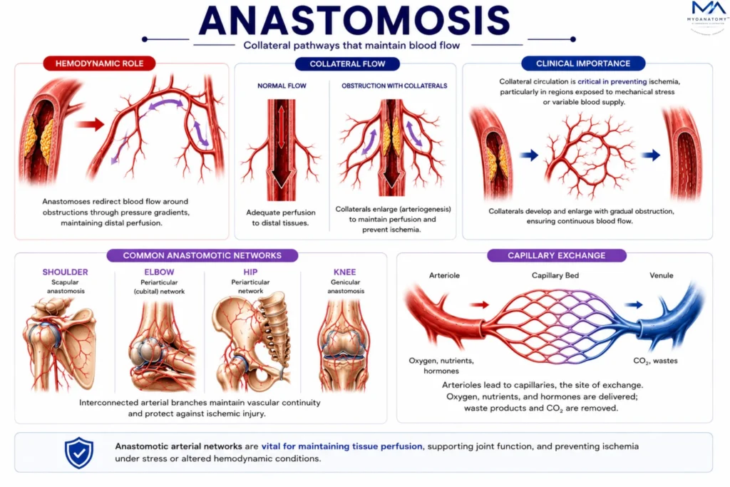
AI Generated llustration MyoAnatomy
Exam Question
Explain how arterial anastomoses preserve tissue perfusion during progressive arterial occlusion, and discuss their hemodynamic and clinical significance in preventing ischemia.
ANATOMY
Scapular Anastomosis
The scapular anastomosis is a collateral arterial network linking branches of the subclavian and axillary arteries, providing an alternative pathway for blood flow to the upper limb when proximal axillary flow is compromised. This network is particularly significant in maintaining perfusion during gradual arterial occlusion or mechanical compression.
Hemodynamically, the scapular anastomosis enables redistribution of blood from the subclavian system to the distal axillary artery via pressure gradients, allowing collateral circulation to sustain limb viability. Over time, these vessels may undergo adaptive enlargement (arteriogenesis), increasing their capacity to compensate for reduced primary arterial flow.
The principal vessels forming this network include the suprascapular artery (from the thyrocervical trunk), the dorsal scapular artery (typically from the subclavian artery), and the circumflex scapular artery (branch of the subscapular artery from the axillary artery). These vessels interconnect within the supraspinous and infraspinous fossae, forming a robust collateral circuit that ensures continuous perfusion of the upper limb.
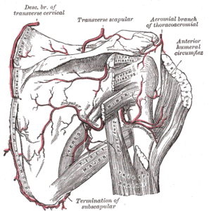
Scapular and Circumflex Arteries (Gray’s Anatomy Plate 521)” – Henry Vandyke Carter, from Henry Gray’s Anatomy of the Human Body, Wikimedia Commons. Public Domain
Exam Question
Explain how the anatomical arrangement of the scapular anastomosis enables collateral circulation between the subclavian and axillary arteries, and analyze its role in maintaining upper limb perfusion during arterial obstruction.
Elbow Anastomosis
The elbow anastomosis is a periarticular arterial network formed by descending collateral branches from the brachial artery and ascending recurrent branches from the radial and ulnar arteries, ensuring continuous blood supply around the elbow joint during movement.
Functionally, this network compensates for transient compression or kinking of the main arterial pathways during flexion and extension of the joint, preserving distal perfusion.
Hemodynamically, pressure gradients facilitate redirection of blood through collateral channels, maintaining flow despite positional or mechanical alterations of primary vessels.
Descending collateral arteries include the superior and inferior ulnar collateral arteries and the radial collateral artery (branch of the profunda brachii), while ascending recurrent arteries include the radial recurrent, anterior and posterior ulnar recurrent, and interosseous recurrent arteries. These vessels form a dense periarticular network surrounding the humeroulnar and humeroradial joints, ensuring vascular continuity and protection against ischemia.
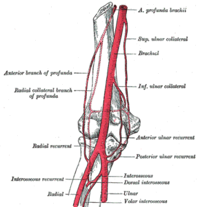
Arteries of the Arm (Gray’s Anatomy Plate 526)” – Henry Vandyke Carter, from Henry Gray’s Anatomy of the Human Body, Wikimedia Commons. Public Domain
Exam Question
Describe the organization of the elbow anastomotic network and analyze how it maintains distal limb perfusion during joint movement and arterial compression.
Genicular Anastomosis
The genicular anastomosis forms a dense vascular network around the knee joint, maintaining perfusion during joint flexion, extension, and weight-bearing.
The principal arteries forming the genicular network include branches of the popliteal artery, with additional contributions from the femoral and anterior tibial arteries.
branches of the popliteal artery
superior medial genicular artery
superior lateral genicular artery
inferior medial genicular artery
inferior lateral genicular artery
Additional contributing arteries
descending genicular artery – branch of the femoral artery
descending branch of the lateral circumflex femoral artery
anterior tibial recurrent artery – branch of the anterior tibial artery
These vessels form a vascular ring surrounding the capsule and ligaments of the knee joint.
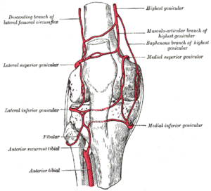
Genicular Anastomosis of the Knee (Gray’s Anatomy Plate 552)” – Henry Vandyke Carter, from Henry Gray’s Anatomy of the Human Body, Wikimedia Commons. Public Domain
Exam Question
Discuss the anatomical organization of the genicular anastomosis, including its principal arterial contributors, and explain its functional importance in maintaining knee perfusion during joint movement and arterial occlusion.
Others Anastomosis
Trochanteric Anastomosis
The trochanteric anastomosis supplies the femoral head and neck and is particularly important for maintaining the vascular supply of the hip joint.
This arterial network is formed by:
ascending branch of the medial circumflex femoral artery
ascending branch of the lateral circumflex femoral artery
superior gluteal artery
inferior gluteal artery
These arteries form a vascular ring around the greater trochanter of the femur and contribute to the retinacular arteries supplying the femoral head.
Cruciate Anastomosis
The cruciate anastomosis lies posterior to the proximal femur and connects arteries of the internal iliac system with those of the femoral system.
The arteries involved include:
medial circumflex femoral artery
lateral circumflex femoral artery
inferior gluteal artery
first perforating artery of the profunda femoris
This network provides collateral circulation between the internal iliac and femoral arterial systems.
Exam Question
Explain how periarticular and proximal femoral arterial anastomoses (genicular, trochanteric, and cruciate) function as collateral networks, and analyze their roles in maintaining lower limb perfusion during joint movement, arterial compression, and pathological vascular compromise.
SUMMARY TABLE