Arterial Supply
MYO CORE
Structural Organization
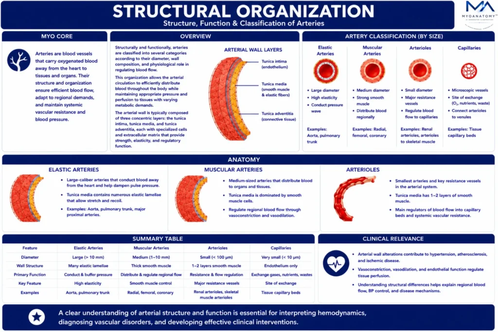
The arterial system forms a hierarchically organized vascular network responsible for transporting oxygenated blood from the heart to peripheral tissues.
OVERVIEW
Structurally and functionally, arteries are classified into several categories according to their diameter, wall composition, and physiological role in regulating blood flow.
This organization allows the arterial circulation to efficiently distribute blood throughout the body while maintaining appropriate pressure and perfusion to tissues with varying metabolic demands.
The arterial wall itself typically consists of three concentric layers: the tunica intima, composed of endothelial cells lining the vessel lumen; the tunica media, containing smooth muscle and elastic fibers that regulate vessel diameter; and the tunica adventitia, a connective tissue layer providing structural support and anchoring the vessel to surrounding tissues.
Variations in the thickness and composition of these layers account for the functional differences between the major categories of arteries.
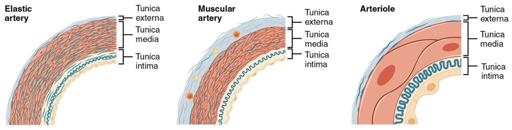
“Muscular and Elastic Artery, Arteriole ( Histological Schematic)” – OpenStax College, via Wikimedia Commons. Licensed under CC BY 3.0
.
Exam Question
Compare the histological organization and biomechanical properties of elastic arteries, muscular arteries, and arterioles, and explain how differences in tunica intima, tunica media, and tunica adventitia determine their specialized roles in arterial pressure regulation, vascular resistance, and tissue perfusion.
ANATOMY
Elastic Artery
Elastic arteries are large-caliber conducting vessels forming the proximal arterial system, specialized for transforming pulsatile ventricular ejection into continuous flow.
Their tunica media contains concentric fenestrated elastic lamellae interspersed with smooth muscle cells, conferring high compliance (ΔV/ΔP) and allowing cyclic energy storage.
During systole, vessel expansion stores elastic energy; during diastole, recoil sustains forward flow and maintains diastolic pressure (Windkessel effect), thereby preserving perfusion of high-demand organs such as the myocardium and brain.
OpenStax College. Anatomy & Physiology (CNX/OpenStax), “Muscular and Elastic Artery, Arteriole.” via Wikimedia Commons. Licensed under CC BY 3.0
Exam Question
Explain how the unique histological architecture of elastic arteries enables the Windkessel effect, and discuss how this adaptation maintains continuous blood flow, dampens pulse pressure, and preserves perfusion of vital organs throughout the cardiac cycle.
Hemodynamically
Hemodynamically, elastic arteries function as capacitance vessels that attenuate pulse pressure, dampen pulsatile energy transmission, and reduce left ventricular afterload while optimizing distal perfusion. They also modulate pulse wave propagation and reflection, limiting systolic pressure augmentation.
Structural integrity depends on elastin-rich extracellular matrix; its degeneration increases arterial stiffness, elevates pulse wave velocity, augments systolic pressure, reduces diastolic pressure, and compromises coronary perfusion.
Exam Question
Explain how the biomechanical properties of elastic arteries influence pulse wave propagation, arterial compliance, ventricular afterload, and distal organ perfusion, and discuss the hemodynamic consequences of age-related arterial stiffening.
Arterial Wall
Endothelial cells regulate vascular homeostasis via shear stress–mediated release of vasoactive mediators (e.g., nitric oxide), while the adventitia contains vasa vasorum supporting the metabolically active vessel wall.
Exam Question
Discuss the structural and functional roles of the tunica intima, tunica media, and tunica adventitia in maintaining arterial wall integrity, vascular homeostasis, and endothelial-mediated regulation of vascular tone under physiological and pathological conditions.
Examples
Representative elastic arteries include – aorta (ascending, arch, thoracic, and abdominal segments), pulmonary trunk, right and left pulmonary arteries, brachiocephalic trunk, common carotid arteries, subclavian arteries, and common iliac arteries.
Their elastic lamellae-rich tunica media enables systolic expansion and diastolic recoil (the Windkessel effect), maintaining continuous blood flow, reducing ventricular afterload, and ensuring stable organ perfusion.
Exam Question
Explain why the aorta, pulmonary trunk, common carotid, subclavian, and common iliac arteries are classified as elastic arteries, and discuss how their elastic lamellae support the Windkessel effect, ventricular unloading, and continuous organ perfusion throughout the cardiac cycle.
Muscular Artery
Muscular arteries are medium-caliber distributing vessels positioned distal to elastic arteries, specialized for regulating regional blood flow.
Their tunica media is composed predominantly of concentric layers of smooth muscle cells with relatively less elastic content, enabling active control of vessel diameter.
Through vasoconstriction and vasodilation, they adjust perfusion according to tissue metabolic demand.
OpenStax College. Anatomy & Physiology (CNX/OpenStax), “Muscular and Elastic Artery, Arteriole.” via Wikimedia Commons. Licensed under CC BY 3.0
Exam Question
Explain how the histological composition of muscular arteries enables precise regulation of regional blood flow, and discuss the physiological importance of smooth muscle–mediated vasoconstriction and vasodilation in tissue perfusion.
Hemodynamically
Hemodynamically, muscular arteries function as flow-distributing vessels rather than pressure buffers, linking central conducting arteries to peripheral resistance vessels. Changes in vascular radius markedly influence blood flow (flow ∝ r⁴), making them critical regulators of regional perfusion.
Their activity is controlled by sympathetic input, circulating hormones, and local metabolic factors, allowing rapid redistribution of blood during physiological states such as exercise and stress
Exam Question
Discuss how changes in muscular artery diameter influence vascular resistance, regional blood flow, and arterial pressure, and explain how neural, hormonal, and local metabolic mechanisms coordinate these hemodynamic responses.
Arterial Wall
The internal elastic lamina provides structural integrity during repeated diameter changes, while dysfunction of smooth muscle tone contributes to impaired perfusion and increased peripheral resistance, playing a key role in hypertension and ischemic pathology.
Exam Question
Describe how the structural organization of the muscular arterial wall, particularly the internal elastic lamina and tunica media, maintains vascular integrity and regulates vessel tone, and explain how their dysfunction contributes to hypertension and ischemic disease.
Examples
Representative muscular arteries include – brachial, radial, ulnar, femoral, popliteal, tibial, coronary, renal, mesenteric, and cerebral arteries.
These medium-sized distributing vessels regulate regional blood flow through smooth muscle–mediated vasoconstriction and vasodilation, ensuring tissue perfusion is precisely matched to the metabolic demands of individual organs and tissues.
Exam Question
Why are the radial, ulnar, brachial, femoral, coronary, renal, mesenteric, and cerebral arteries classified as muscular arteries, and how does their structure support regional blood flow distribution and organ-specific perfusion?
Arteriols
Arterioles are the smallest branches of the arterial system and function as the primary resistance vessels regulating blood flow into capillary networks.
Their walls consist of a thin tunica media with one to a few layers of smooth muscle cells, yet these cells exert a disproportionate influence on vascular resistance and systemic blood pressure.
OpenStax College. Anatomy & Physiology (CNX/OpenStax), “Muscular and Elastic Artery, Arteriole.” via Wikimedia Commons. Licensed under CC BY 3.0
Exam Question
Explain how the structural organization of arterioles enables them to function as the principal resistance vessels, and discuss their role in regulating systemic vascular resistance, arterial pressure, and capillary perfusion.
Hemodynamically
Hemodynamically, arterioles are the principal determinants of total peripheral resistance, where small changes in luminal radius produce large changes in flow (flow ∝ r⁴). Through vasoconstriction and vasodilation, they regulate tissue perfusion and redistribute cardiac output according to metabolic demand.
Their tone is tightly controlled by sympathetic nervous activity, circulating hormones, and local metabolic factors (e.g., hypoxia, CO₂, H⁺), enabling precise, moment-to-moment regulation of blood flow.
Exam Question
Discuss how arteriolar vasoconstriction and vasodilation regulate total peripheral resistance and regional blood flow, and explain how neural, hormonal, and local metabolic mechanisms coordinate these hemodynamic responses.
Arterial Wall
Functionally, arterioles serve as the critical interface between macrocirculation and microcirculation, controlling pressure drop across the vascular system and protecting capillary beds from excessive pressure.
Dysregulation of arteriolar tone leads to altered peripheral resistance and is central to the pathophysiology of hypertension, shock, and impaired tissue perfusion
Exam Question
Describe how the arteriolar wall maintains the transition between macrocirculation and microcirculation, and explain how dysregulation of arteriolar tone contributes to hypertension, shock, and impaired tissue perfusion.
Examples
Representative arterioles examples are afferent and efferent renal arterioles, coronary arterioles, cerebral arterioles, cutaneous arterioles, skeletal muscle arterioles, and mesenteric arterioles.
These terminal branches of the arterial system regulate blood entry into capillary beds through vasoconstriction and vasodilation, making them the principal resistance vessels controlling tissue perfusion and systemic vascular resistance.
Exam Question
Why are afferent and efferent renal, coronary, cerebral, skeletal muscle, mesenteric, and cutaneous arterioles classified as resistance vessels, and how does regulation of their diameter determine regional perfusion, capillary pressure, and systemic vascular resistance?
Capillaries
Capillaries are the smallest and most numerous vessels, forming the microvascular interface between arterial inflow and venous return and serving as the primary site of exchange between blood and tissues.
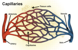
Source: National Cancer Institute, National Institutes of Health (NIH). Capillaries illustration. Available via Wikimedia Commons. Public Domain (U.S. Federal Government Work).
Exam Question
Explain how the ultrastructural organization of capillaries optimizes diffusion, filtration, and transcytosis, and discuss how these mechanisms maintain tissue oxygenation, nutrient delivery, and metabolic homeostasis.
Hemodynamically
Functionally, capillaries facilitate the exchange of gases, nutrients, metabolites, and signaling molecules via diffusion, filtration, and transcytosis, governed by concentration gradients and Starling forces. Their organization into capillary beds allows dynamic regulation of flow through precapillary sphincters and local metabolic control, ensuring preferential perfusion of metabolically active tissues.
Within the musculoskeletal system, capillary networks support oxidative metabolism, tissue repair, and adaptation to mechanical load, with capillary density closely correlating with metabolic demand and increasing in response to endurance training. Impairment of capillary function disrupts tissue oxygenation and nutrient delivery, contributing to ischemia and impaired healing.
Exam Question
Discuss how capillary exchange is regulated by Starling forces, concentration gradients, and precapillary sphincters, and explain how alterations in these mechanisms contribute to edema, ischemia, and impaired tissue repair.
Arterial Wall
Structurally, they consist of a single layer of endothelial cells supported by a basement membrane, minimizing diffusion distance and optimizing exchange efficiency.
Exam Question
Describe how the single endothelial cell layer, basement membrane, and endothelial permeability regulate selective molecular exchange, and explain how disruption of capillary wall integrity contributes to inflammation, vascular leakage, and organ dysfunction.
Examples
Representative arterioles examples are continuous capillaries (skeletal muscle, skin, lungs, and central nervous system), fenestrated capillaries (kidneys, endocrine glands, and intestinal villi), and sinusoidal capillaries (liver, spleen, and bone marrow).
Their structural specialization optimizes exchange according to the metabolic and functional demands of each tissue.
Exam Question
Compare continuous, fenestrated, and sinusoidal capillaries, and explain how their structural specializations optimize tissue-specific exchange while maintaining organ function and homeostasis.
SUMMARY TABLE