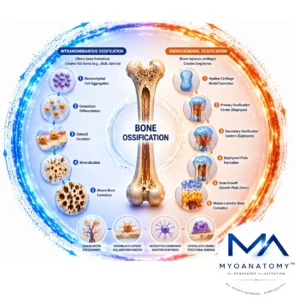
Bone Ossification
MYO CORE
Endochondral Ossification
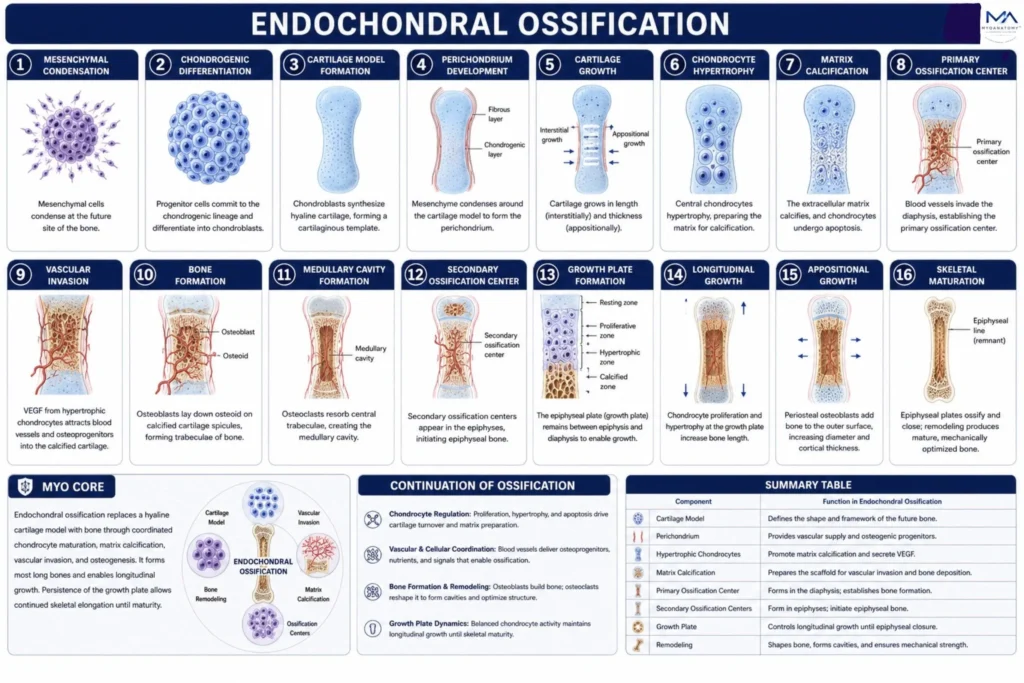
Endochondral ossification is the process by which a hyaline cartilage model is progressively replaced by bone tissue. Through coordinated chondrocyte maturation, vascular invasion, matrix calcification, and osteogenic differentiation, it forms most bones of the appendicular skeleton and enables longitudinal growth. Persistence of the epiphyseal plate maintains growth potential until skeletal maturity, supporting skeletal enlargement, biomechanical adaptation, and structural maturation.
OVERVIEW
Cartilage Model development
Endochondral ossification is initiated by the differentiation of mesenchymal stem cells into chondroblasts, which synthesize a hyaline cartilage template that replicates the shape of the future bone. This cartilage model serves as a transient structural scaffold guiding subsequent bone formation.
Cartilage growth occurs via two complementary mechanisms:
Interstitial growth, driven by chondrocyte proliferation within the matrix, leading to internal expansion, and appositional growth, involving the addition of new cartilage layers at the surface from the perichondrium. Together, these processes enable controlled enlargement and morphological refinement of the developing skeletal element.
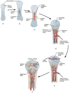
“608 Endochondral Ossification” – Pearson Scott Foresman via Wikimedia Commons. Public Domain.
ANATOMY
Continuation of Ossification
Following establishment of the cartilage model, endochondral ossification progresses through a tightly regulated sequence of chondrocyte proliferation, hypertrophy, matrix calcification, vascular invasion, osteogenic differentiation, and progressive replacement of cartilage by bone.
Primary and secondary ossification centers coordinate the formation of the diaphysis and epiphyses, while the epiphyseal growth plate maintains longitudinal growth until closure.
This process transforms a temporary cartilaginous scaffold into a mature, vascularized, mechanically competent skeletal structure.
Exam Question
How do primary and secondary ossification centers coordinate cartilage replacement, skeletal growth, and the formation of mature bone during endochondral ossification?
1. Mesenchymal Condensation
Mesenchymal condensation represents the earliest stage of endochondral ossification and establishes the anatomical blueprint of the future skeletal element.
Mesenchymal stem cells migrate, proliferate, and aggregate within highly vascular embryonic connective tissue, forming localized cellular condensations that define the precise shape, size, orientation, and location of the developing bone.
Increased cell-to-cell interactions, extracellular matrix remodeling, and activation of osteochondrogenic signaling pathways create a specialized microenvironment that promotes commitment toward the chondrogenic lineage.
This condensation serves as the developmental template from which the cartilaginous model of the future bone will subsequently arise.
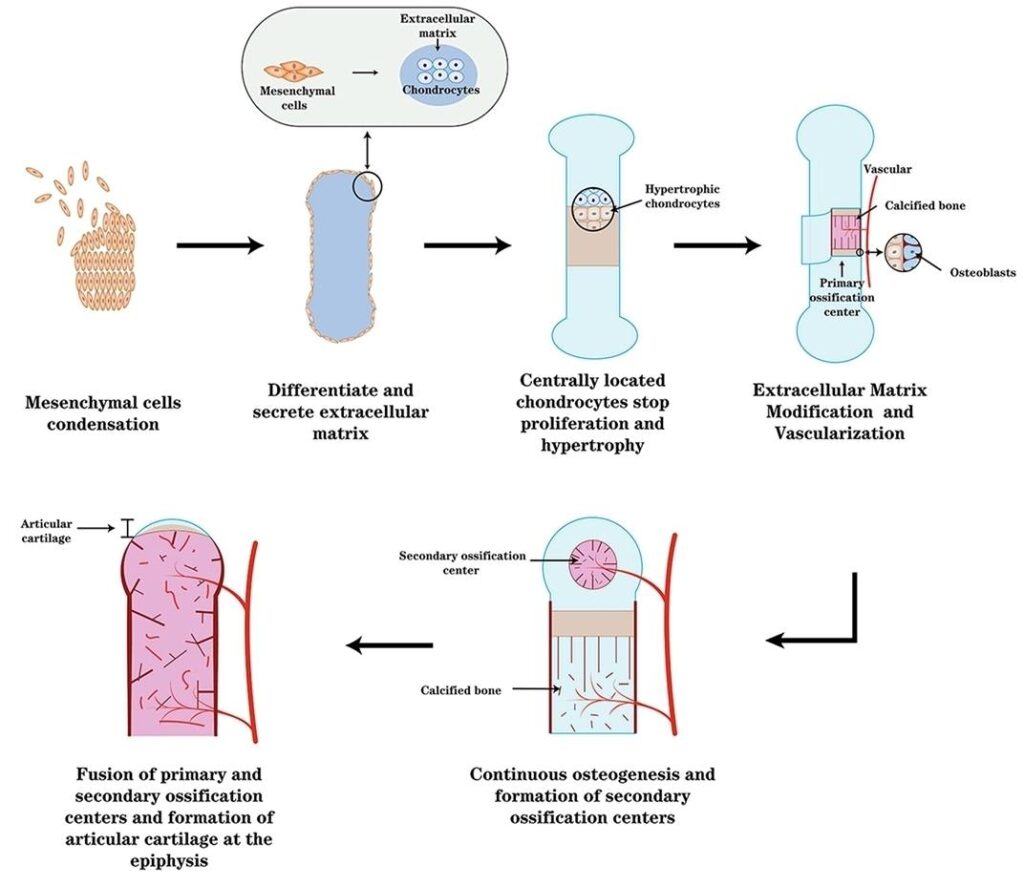
“Formation of Endochondral Ossification and Articular Cartilage” – Zhangpeng Su et al., via Wikimedia Commons. Licensed under CC BY 4.0.
Exam Question
How do mesenchymal cell aggregation, extracellular matrix remodeling, and embryonic signaling pathways interact to establish the developmental framework required for formation of the future endochondral skeletal element?
2. Chondrogenic Differentiation
Following mesenchymal condensation, progenitor cells commit to the chondrogenic lineage and differentiate into chondroblasts, the specialized cells responsible for cartilage formation.
This transition is driven primarily by SOX9, together with SOX5 and SOX6, which activate genes required for synthesis of cartilage extracellular matrix. As differentiation progresses, chondroblasts begin producing type II collagen and proteoglycan-rich matrix, establishing the molecular foundation of the future cartilage model.
This stage represents the critical developmental shift from undifferentiated embryonic mesenchyme to a dedicated cartilage-forming tissue that will subsequently serve as the template for endochondral bone formation.
Exam Question
Why is SOX9-mediated chondrogenic differentiation considered the pivotal commitment step of endochondral ossification, and how would disruption of this process affect formation of the future skeletal template?
3. Cartilage Model Formation
As chondroblasts proliferate and secrete increasing amounts of cartilage matrix, a hyaline cartilage model of the future bone is established.
This cartilaginous template closely replicates the size, shape, and overall morphology of the developing skeletal element, providing a temporary structural framework for subsequent ossification. Embedded within lacunae, chondrocytes maintain and expand the matrix while preserving the architectural integrity of the model.
The cartilage model serves as the indispensable scaffold upon which all subsequent stages of endochondral bone formation depend.
Exam Question
How does formation of the hyaline cartilage model establish the structural and developmental template required for subsequent vascular invasion, ossification center formation, and mature bone development?
4. Perichondrium Development
As the cartilage model enlarges, the surrounding mesenchymal tissue condenses to form the perichondrium, a specialized connective tissue sheath that envelops the developing cartilage. This structure provides mechanical support, facilitates nutrient diffusion to the avascular cartilage, and serves as a reservoir of progenitor cells.
Progressive vascularization and osteogenic signaling within the inner perichondrial layer prepare it for subsequent transformation into the periosteum, thereby linking cartilage development to future bone formation.
The perichondrium therefore functions as a critical regulatory interface between the growing cartilage template and the emerging skeletal tissue.
Exam Question
How does perichondrium development support cartilage growth while simultaneously establishing the cellular and molecular conditions necessary for subsequent osteogenesis during endochondral ossification?
5.Cartilage Growth
Following formation of the cartilage model, the developing template enlarges through both interstitial and appositional growth. Interstitial growth occurs through chondrocyte proliferation and matrix expansion from within the cartilage, while appositional growth results from the addition of new cartilage by chondroblasts located in the inner layer of the perichondrium.
Together, these complementary mechanisms increase the length, diameter, and overall mass of the cartilage model while preserving its structural organization. This stage establishes the size and morphology required for subsequent hypertrophy, vascular invasion, and ossification.
Exam Question
How do interstitial and appositional growth mechanisms coordinate expansion of the cartilage model while maintaining the structural integrity necessary for endochondral ossification?
6. Chondrocytes Hypertrophy
Chondrocyte hypertrophy marks the transition from cartilage growth to ossification. Chondrocytes within the central region of the cartilage model undergo marked enlargement, accompanied by profound metabolic and genetic changes that prepare the matrix for mineralization.
Hypertrophic chondrocytes modify the extracellular matrix, promote its calcification, and secrete angiogenic factors-most notably VEGF-that stimulate vascular invasion.
Consequently, these cells function not merely as structural components of cartilage but as critical regulators that orchestrate the transition from a cartilaginous template to developing bone.
Exam Question
How do hypertrophic chondrocytes coordinate matrix calcification, angiogenesis, and the transition from cartilage growth to bone formation during endochondral ossification?
7. Matrix Calcification
Matrix calcification occurs when the extracellular cartilage matrix surrounding hypertrophic chondrocytes becomes progressively mineralized through deposition of calcium and phosphate salts.
As calcification advances, diffusion of oxygen and nutrients through the matrix is markedly reduced, leading to chondrocyte apoptosis and degeneration of the central cartilage tissue.
This process converts the flexible cartilage scaffold into a rigid mineralized framework capable of supporting vascular invasion and subsequent bone deposition. Matrix calcification therefore represents the critical transitional stage that prepares cartilage for replacement by skeletal tissue.
Exam Question
How does matrix calcification transform the cartilage template into a mineralized scaffold capable of supporting vascular invasion, chondrocyte apoptosis, and subsequent bone formation during endochondral ossification?
8. Primary Ossification Center
The primary ossification center develops within the diaphysis following matrix calcification and vascular invasion.
As blood vessels penetrate the calcified cartilage, they introduce osteoprogenitor cells, osteoblast precursors, osteoclast precursors, and regulatory growth factors into the developing skeletal tissue.
Osteoblasts begin depositing osteoid onto remnants of calcified cartilage, creating the first trabeculae of primary bone.
This stage marks the initiation of true bone formation and establishes the principal center from which ossification expands throughout the developing shaft.
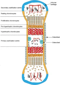
“Schematic of Endochondral Ossification and Formation of Primary and Secondary Ossification Centers” – Javaheri B. et al. (2018) via Wikimedia Commons. Licensed under CC BY 4.0.
Exam Question
How does formation of the primary ossification center coordinate vascular invasion, osteogenic recruitment, and replacement of calcified cartilage by developing bone during endochondral ossification?
9. Vascular Invasion
Vascular invasion occurs when blood vessels penetrate the calcified cartilage matrix and enter the developing primary ossification center.
Driven largely by VEGF released from hypertrophic chondrocytes, invading vessels transport osteoprogenitor cells, osteoblast precursors, osteoclast precursors, nutrients, minerals, and regulatory growth factors into previously avascular cartilage.
Beyond nutrient delivery, this process establishes the cellular and molecular environment necessary for cartilage resorption, osteogenesis, marrow development, and continued skeletal growth. Vascular invasion therefore serves as the pivotal event that transforms a calcified cartilage template into a metabolically active bone-forming organ.
Exam Question
How does VEGF-mediated vascular invasion coordinate cellular recruitment, cartilage replacement, osteogenesis, and marrow development during endochondral ossification?
10. Bone Formation
Bone formation begins when osteoprogenitor cells delivered through invading blood vessels differentiate into osteoblasts within the primary ossification center. These osteoblasts deposit osteoid onto remnants of calcified cartilage, creating the first trabeculae of primary bone. Through continuous matrix synthesis and mineralization, the temporary cartilage scaffold is progressively replaced by developing skeletal tissue.
This stage represents the fundamental transition from a cartilaginous template to true bone and establishes the structural framework for subsequent skeletal growth, remodeling, and maturation.
Exam Question
How do osteoblast differentiation, osteoid deposition, and cartilage replacement coordinate the transition from a calcified cartilage template to developing bone during endochondral ossification?
11. Medullary Cavity Formation
Medullary cavity formation occurs as osteoclasts progressively resorb central trabeculae within the primary ossification center, creating an expanding internal cavity within the diaphysis.
This remodeling process removes excess primary bone while preserving structural stability, allowing the developing shaft to become lighter, mechanically efficient, and capable of housing bone marrow.
Simultaneously, the newly formed cavity accommodates hematopoietic tissue, blood vessels, and marrow stromal cells, transforming the developing bone into both a structural and metabolic organ. This stage is essential for establishing the characteristic internal architecture of mature long bones.
Exam Question
How does osteoclastic remodeling within the primary ossification center generate the medullary cavity while preserving skeletal strength and enabling marrow development during endochondral ossification?
12. Secondary Ossification Center
Secondary ossification centers develop within the epiphyses when blood vessels invade the cartilaginous ends of the bone and introduce osteogenic precursor cells. Similar to the primary ossification center, osteoblasts begin depositing osteoid onto calcified cartilage remnants, initiating bone formation within the epiphysis.
Unlike the diaphysis, however, a medullary cavity does not develop, allowing the epiphyseal region to retain its trabecular architecture.
The emergence of secondary ossification centers separates the epiphysis from the diaphysis and establishes the foundation for the future growth plate.
“Schematic of Endochondral Ossification and Formation of Primary and Secondary Ossification Centers” – Javaheri B. et al. (2018) via Wikimedia Commons. Licensed under CC BY 4.0.
Exam Question
How do secondary ossification centers coordinate epiphyseal bone formation while preserving the structural and developmental conditions necessary for continued longitudinal growth?
13. Growth Plate Formation
Growth plate formation occurs when a layer of hyaline cartilage persists between the primary ossification center of the diaphysis and the secondary ossification centers of the epiphyses. Rather than undergoing immediate ossification, this cartilage becomes organized into distinct cellular zones that regulate chondrocyte proliferation, hypertrophy, calcification, and replacement by bone.
The epiphyseal plate therefore functions as the principal growth center of the developing long bone, maintaining a balance between cartilage production and ossification that permits continued skeletal elongation while preserving structural continuity between the epiphysis and diaphysis.
Exam Question
How does growth plate organization regulate the balance between cartilage proliferation and ossification to sustain longitudinal skeletal growth during development?
14. Longitudinal growth
Longitudinal growth occurs at the epiphyseal plate through the continuous proliferation, hypertrophy, calcification, and ossification of chondrocytes. As new cartilage is produced on the epiphyseal side and simultaneously replaced by bone on the diaphyseal side, the length of the bone progressively increases while the growth plate itself is maintained.
This highly regulated process enables skeletal elongation throughout childhood and adolescence and continues until epiphyseal closure at skeletal maturity. Longitudinal growth therefore represents the principal mechanism by which long bones achieve their final length and anatomical proportions.
Exam Question
How does the coordinated balance between chondrocyte proliferation, hypertrophy, and ossification within the epiphyseal plate drive longitudinal skeletal growth while maintaining growth plate integrity?
15. Appositional Growth
Appositional growth increases the diameter and cortical thickness of developing bone through osteoblastic activity within the periosteum.
As osteoblasts deposit new bone matrix along the external surface, simultaneous endosteal remodeling preserves appropriate cortical proportions and medullary cavity dimensions. This coordinated process allows the growing skeleton to increase in width and mechanical strength while adapting to progressively greater functional and biomechanical demands.
Appositional growth therefore complements longitudinal growth by optimizing skeletal robustness, load-bearing capacity, and structural stability.
Exam Question
How does coordinated periosteal bone deposition and endosteal remodeling regulate appositional growth while maintaining skeletal strength and architectural efficiency during development?
16. Skeletal Maturation
Skeletal maturation represents the final stage of endochondral ossification, during which bone growth progressively ceases as the epiphyseal plate undergoes complete ossification and is replaced by the epiphyseal line.
Concurrent remodeling of cortical and trabecular compartments optimizes skeletal architecture according to mechanical demands, producing the mature balance between strength, mass, and metabolic function.
Although longitudinal growth ends, the skeleton remains a dynamic tissue capable of continuous remodeling, repair, and adaptation throughout life. This stage marks the transition from a growing skeletal framework to a fully mature and mechanically optimized skeletal system.
Exam Question
How do epiphyseal closure and lifelong skeletal remodeling collectively transform the developing skeleton into a mature, mechanically optimized, and metabolically active organ?
SUMMARY TABLE