Bone Ossification
MYO CORE
Intramembraneous Ossification
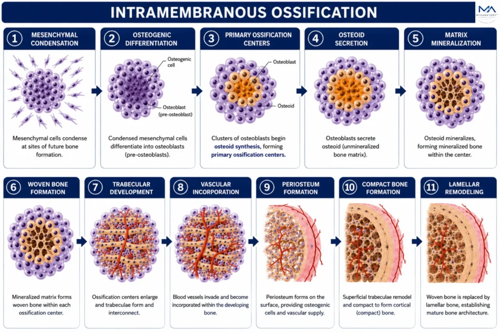
Intramembranous ossification is the direct formation of bone from embryonic mesenchymal tissue without a cartilaginous precursor. Mesenchymal stem cells differentiate into osteoblasts that synthesize osteoid, initiate mineralization, and establish primary trabecular bone. Subsequent remodeling produces mature lamellar bone, enabling the development of most flat bones of the skull, portions of the facial skeleton, and the clavicle while supporting skeletal growth, structural integrity, and mineral homeostasis.
OVERVIEW
Initiation of Osteogenesis
It underlies embryonic development, postnatal growth, fracture repair, and lifelong skeletal maintenance, ensuring mechanical stability, protection, and calcium–phosphate homeostasis.
This process is mediated by integrated activity of osteogenic lineage cells within a vascularized environment, where osteoblasts deposit a collagen-rich matrix that subsequently mineralizes, conferring rigidity and load-bearing capacity, while osteocytes coordinate adaptive responses and osteoclasts enable structural renewal.
Ossification occurs via intramembranous and endochondral pathways, which differ in developmental sequence but converge in the formation of lamellar bone optimized for mechanical function. Throughout life, ossification remains dynamically regulated by mechanical forces, hormonal control, and molecular signaling, ensuring continuous adaptation, structural integrity, and metabolic balance of bone tissue.
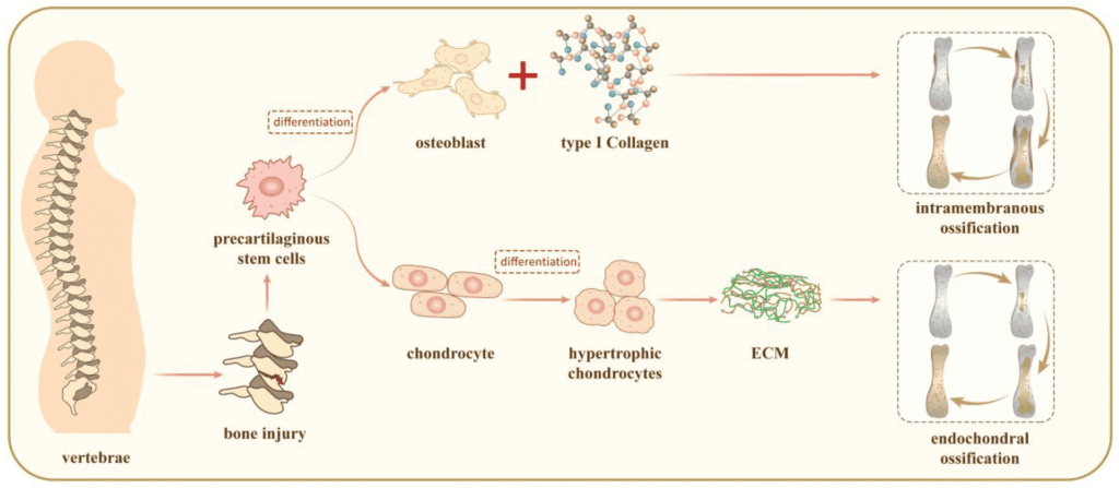
“Intramembranous ossification and endochondral ossification” – Ge R., Liu C., Zhao Y., Wang K., Wang X via Wikimedia Commons. Licensed under CC BY 4.0.
Exam Question
Explain the molecular mechanisms that govern mesenchymal stem cell commitment to the osteogenic lineage, including the role of the RUNX2, Osterix, BMP, Wnt/b-catenin signaling in the initiating bone formation.
ANATOMY
1. Mesenchymal Condensation
Mesenchymal condensation is the initiating morphogenetic event of intramembranous ossification, in which mesenchymal stem cells aggregate within highly vascular embryonic connective tissue to define the future sites of membrane bone formation.
Increased cellular density enhances cell-to-cell communication and activates osteogenic signaling pathways, especially BMP, RUNX2, and Wnt/β-catenin, driving local mesenchyme toward an osteogenic fate.
This stage establishes the spatial blueprint, cellular foundation, and developmental microenvironment required for direct bone formation without a cartilage precursor.
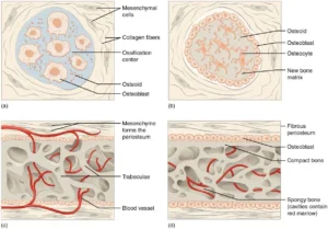
“Intramembraneous Ossification” by Open Stax College, from Anatomy&Physiology (OpenStax), via Wikimedia Commons. Licenswed under CC BY 3.0
Exam Question
How do mesenchymal condensation, vascular signaling, and osteogenic pathway activation establish the developmental foundation for direct bone formation during intramembranous ossification?
2. Osteogenic Differentiation
Osteogenic differentiation is the process by which condensed mesenchymal stem cells transform into osteoprogenitor cells and subsequently mature osteoblasts.
Driven primarily by RUNX2, Osterix (SP7), and BMP signaling, this stage initiates the genetic and cellular program required for bone formation.
As differentiation progresses, cells acquire the capacity to synthesize osteoid, regulate mineralization, and establish ossification centers. This transition represents the definitive conversion of embryonic mesenchyme into specialized bone-forming tissue and marks the beginning of active osteogenesis.
“Intramembraneous Ossification” by Open Stax College, from Anatomy&Physiology (OpenStax), via Wikimedia Commons. Licenswed under CC BY 3.0
Exam Question
How do RUNX2, Osterix, and BMP signaling coordinate the differentiation of mesenchymal stem cells into functional osteoblasts capable of initiating bone formation?
3. Ossification Centers
Ossification centers are the first focal sites of active bone formation within condensed mesenchymal tissue. Following osteogenic differentiation, clusters of osteoblasts accumulate and initiate localized osteoid synthesis, creating regions where mineralized bone development begins.
These centers function as biological hubs of osteogenesis, coordinating matrix production, mineralization, and outward skeletal expansion.
As multiple ossification centers enlarge and fuse, they progressively transform embryonic connective tissue into a continuous network of developing bone, establishing the foundational architecture of future membrane bones.
“Intramembraneous Ossification” by Open Stax College, from Anatomy&Physiology (OpenStax), via Wikimedia Commons. Licenswed under CC BY 3.0
Exam Question
How do multiple intramembranous ossification centers coordinate localized osteogenesis and subsequent fusion to establish the structural framework of developing membrane bones?
4.Osteoid Secretion
Osteoid secretion begins when osteoblasts actively synthesize and release the organic extracellular matrix that serves as the foundation of future bone tissue.
This matrix consists predominantly of type I collagen, together with osteocalcin, osteonectin, proteoglycans, and other non-collagenous proteins that regulate matrix organization and mineral deposition.
Osteoid establishes the structural scaffold upon which mineralization occurs, defining the architecture, strength, and biological properties of developing bone. Without osteoid formation, mineralized skeletal tissue cannot develop.
“Intramembraneous Ossification” by Open Stax College, from Anatomy&Physiology (OpenStax), via Wikimedia Commons. Licenswed under CC BY 3.0
Exam Question
Why is osteoid secretion considered the essential biosynthetic foundation of bone formation, and how does its molecular composition influence subsequent skeletal mineralization?
5. Matrix Mineralisation
Matrix mineralization is the process by which the osteoid matrix becomes transformed into rigid bone through the controlled deposition of calcium and phosphate as hydroxyapatite crystals.
These crystals nucleate along collagen fibrils and progressively infiltrate the organic matrix, increasing its density, stiffness, and mechanical strength. Precise regulation of mineral deposition is essential to achieve the balance between rigidity and fracture resistance that characterizes healthy bone.
This stage converts biologically synthesized osteoid into functional mineralized skeletal tissue capable of supporting mechanical loads.
“Intramembraneous Ossification” by Open Stax College, from Anatomy&Physiology (OpenStax), via Wikimedia Commons. Licenswed under CC BY 3.0
Exam Question
How does hydroxyapatite crystal deposition transform osteoid into mechanically competent bone, and why is precise regulation of mineralization essential for skeletal function?
6. Woven Bone Formation
Following mineralization, newly formed bone appears as woven bone, the first immature skeletal tissue produced during intramembranous ossification.
Woven bone is characterized by rapidly deposited matrix, randomly oriented collagen fibers, high cellularity, and abundant osteocytes embedded within lacunae. Although mechanically weaker than mature lamellar bone, its rapid formation provides immediate structural continuity and supports ongoing skeletal expansion.
This temporary framework serves as the precursor from which the mature architecture of bone subsequently develops through remodeling.
“Intramembraneous Ossification” by Open Stax College, from Anatomy&Physiology (OpenStax), via Wikimedia Commons. Licenswed under CC BY 3.0
Exam Question
Why is woven bone ideally suited for rapid skeletal formation, and how do its structural characteristics necessitate subsequent remodeling into lamellar bone?
7. Trabecular development
Trabecular development occurs as newly formed woven bone undergoes structural expansion and reorganization into an interconnected three-dimensional network of bony trabeculae. Through continuous osteoblastic deposition and architectural remodeling, individual bone spicules enlarge, fuse, and align along regions of functional stress, creating primary spongy bone.
This highly vascular framework maximizes biomechanical efficiency by distributing forces throughout the developing skeleton while minimizing skeletal mass.
Simultaneously, the spaces between trabeculae accommodate vascular ingrowth and future marrow formation, transforming primitive bone into a dynamic structural and metabolic organ. As a result, trabecular architecture establishes the internal framework upon which mature skeletal strength, adaptation, and remodeling depend.
“Intramembraneous Ossification” by Open Stax College, from Anatomy&Physiology (OpenStax), via Wikimedia Commons. Licenswed under CC BY 3.0
Exam Question
How does trabecular organization integrate biomechanical optimization, vascular accommodation, and skeletal growth to establish the functional architecture of developing bone?
8. Vascular Incorporation
Vascular incorporation begins when blood vessels penetrate the spaces between developing trabeculae and establish a functional vascular network throughout the immature bone.
Driven largely by VEGF-mediated angiogenesis, this process supplies oxygen, nutrients, mineral substrates, regulatory growth factors, and osteogenic precursor cells required for continued skeletal development.
Beyond simple nutrient delivery, invading vessels function as biological signaling centers that coordinate osteoblast activity, matrix maturation, tissue remodeling, and future marrow formation. Vascular integration therefore transforms primitive trabecular bone into a metabolically active, continuously growing, and dynamically remodeling skeletal organ.
“Intramembraneous Ossification” by Open Stax College, from Anatomy&Physiology (OpenStax), via Wikimedia Commons. Licenswed under CC BY 3.0
Exam Question
How does VEGF-mediated vascular incorporation coordinate osteogenic recruitment, metabolic support, marrow development, and skeletal maturation during intramembranous ossification?
9. Periosteum Formation
Periosteum formation occurs when peripheral mesenchymal cells surrounding the developing bone condense into a specialized connective tissue membrane composed of an outer fibrous layer and an inner osteogenic layer.
This structure establishes the primary reservoir of osteoprogenitor cells and osteoblasts responsible for appositional bone growth. Beyond its developmental role, the periosteum becomes a critical regulatory interface that supports vascular supply, skeletal remodeling, fracture repair, and lifelong bone maintenance.
Its formation therefore transforms the developing bone from a simple trabecular framework into a self-renewing organ capable of growth, adaptation, and regeneration.
“Intramembraneous Ossification” by Open Stax College, from Anatomy&Physiology (OpenStax), via Wikimedia Commons. Licenswed under CC BY 3.0
Exam Question
How does periosteum formation establish the osteogenic, vascular, and regenerative capacity required for skeletal growth, remodeling, and lifelong maintenance?
10. Compact Bone Formation
Compact bone formation occurs when superficial trabeculae of primary spongy bone undergo progressive remodeling and condensation to produce dense cortical bone at the outer surface of the developing skeleton.
Through coordinated osteoblastic matrix deposition and osteoclastic refinement, trabecular bone is reorganized into a highly compact structure characterized by increased mineral density and reduced porosity.
This transformation generates the rigid external framework of bone, providing mechanical strength, structural stability, and resistance to compression, bending, and torsional forces.
Compact bone formation therefore establishes the protective and load-bearing architecture required for mature skeletal function.
“Intramembraneous Ossification” by Open Stax College, from Anatomy&Physiology (OpenStax), via Wikimedia Commons. Licenswed under CC BY 3.0
Exam Question
How does the remodeling of primary trabecular bone into compact cortical bone optimize skeletal strength, structural stability, and biomechanical performance during intramembranous ossification?
11. Lamellar Remodeling
Lamellar remodeling is the final maturation stage of intramembranous ossification, during which immature woven bone is progressively replaced by highly organized lamellar bone.
Through tightly coupled osteoclastic resorption and osteoblastic deposition, collagen fibers become arranged in parallel lamellae aligned with mechanical stress lines, producing a structurally optimized skeletal architecture. Simultaneously, mature trabecular networks and cortical organization are refined to maximize strength, durability, and biomechanical efficiency.
This continuous remodeling process transforms rapidly formed developmental bone into a mature skeleton capable of lifelong adaptation, repair, and functional loading.
“Intramembraneous Ossification” by Open Stax College, from Anatomy&Physiology (OpenStax), via Wikimedia Commons. Licenswed under CC BY 3.0
Exam Question
How does coordinated osteoclast–osteoblast remodeling transform immature woven bone into mechanically optimized lamellar bone capable of lifelong adaptation and structural maintenance?
SUMMARY TABLE