Although the majority of articulations within the skull are fibrous sutures and the cranial base contains temporary cartilaginous joints (synchondroses), a small number of true synovial joints exist within the craniofacial skeleton. These joints represent the only freely movable articulations of the skull, permitting localized motion necessary for several essential physiological processes.
Temporomandibular joint
CORE
Overview
Structurally, synovial joints share the classical features characteristic of diarthrodial joints:
articular surfaces covered by cartilage
a synovial cavity containing lubricating synovial fluid
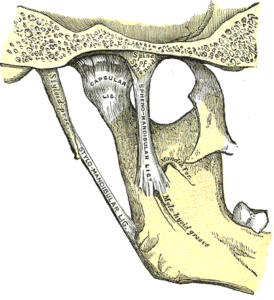
a fibrous articular capsule
a synovial membrane lining the capsule
accessory ligaments and stabilizing structures
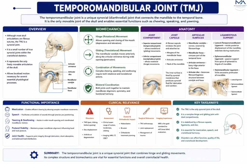
However, synovial joints of the skull show important specialized adaptations reflecting their functional roles in mastication and auditory mechanics.
Within the skull, synovial joints occur primarily in two anatomical systems:
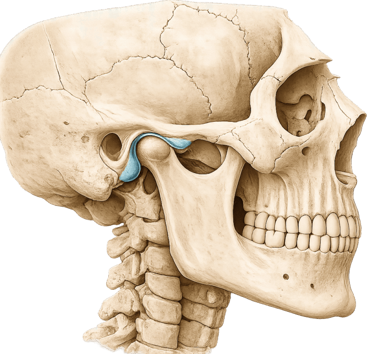
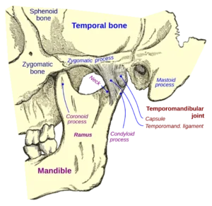
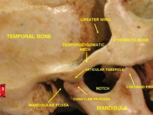
1.Masticatory apparatus – the temporomandibular joint
2.Auditory apparatus – synovial joints between the auditory ossicles
Despite their small number, these joints play a critical role in integrating skeletal mechanics, neuromuscular coordination, and sensory function within the head.


ANATOMY
Anatomical Classification
The temporomandibular joint is the largest and most complex synovial joint of the skull. It represents the articulation between the mandible, the only movable bone of the skull, and the temporal bone.
Functionally and structurally, the TMJ is classified as a modified hinge joint (ginglymoarthrodial joint) because it combines:
Rotational (hinge) movement
Translational (gliding) movement
This dual mechanism allows the mandible to perform complex movements required for chewing, speaking, swallowing, and facial expression.
Unlike most synovial joints of the body, the TMJ possesses a fibrocartilaginous articular surface, which provides greater resistance to mechanical stress and compressive forces generated during mastication.
Exam Question
Explain why the temporomandibular joint is classified as a modified hinge (ginglymoarthrodial) joint, and describe how its dual movement pattern enables functional mandibular activity.
Articulating Surfaces
The temporomandibular joint is formed by articulation between 3 primary anatomical structures:
mandibular condyle (head of the mandible) – an elliptical bony prominence oriented mediolaterally. It articulates with the mandibular fossa,
mandibular fossa of the temporal bone – shallow depression on the inferior surface of the temporal bone.
articular tubercle (articular eminence) of the temporal bone
During mandibular movement, the condyle glides along the articular tubercle, particularly during wide opening of the mouth.
Exam Question
Identify the three primary articulating components of the temporomandibular joint and explain how their morphology facilitates combined rotational and translational movements.
Articular Cartilage
A unique feature of the TMJ is that the articular surfaces are covered by fibrocartilage rather than hyaline cartilage.
Fibrocartilage provides several functional advantages:
greater resistance to shear forces
enhanced durability under repeated mechanical stress
improved ability to remodel in response to loading.
These characteristics are essential for a joint that experiences frequent and powerful forces during chewing.
Exam Question
Why are the articular surfaces of the temporomandibular joint covered by fibrocartilage instead of hyaline cartilage, and how does this adaptation relate to functional loading?
Articular Disk
A defining structural component of the TMJ is the articular disc, a dense fibrocartilaginous structure interposed between the mandibular condyle and the temporal bone.
Morphology
The disc is biconcave, with: a thin central zone thicker anterior and posterior bands
Posteriorly the disc is attached to the retrodiscal tissue, a highly vascular and innervated connective tissue region.
Anteriorly the disc attaches to the lateral pterygoid muscle, allowing muscular control of disc position.
Exam Question
Describe the morphology and attachments of the articular disc of the temporomandibular joint, and explain its role in load distribution and joint stability.
Joint Compartment
The articular disc divides the joint cavity into two functionally distinct compartments:
Superior joint compartment
allows translational (gliding) movements between the disc and temporal bone.
responsible for mandibular protrusion wide opening of the mouth
Inferior joint compartment
allows rotational (hinge) movements between the condyle and the disc.
responsible for: early phase of mouth opening and closing
Exam Question
Explain the functional division of the temporomandibular joint cavity into superior and inferior compartments, and correlate each with specific mandibular movements.
Joint Capsule
The temporomandibular joint is enclosed by a fibrous capsule that attaches to:
the margins of the mandibular fossa
the articular tubercle
the neck of the mandible
The inner surface of the capsule is lined by a synovial membrane, which produces synovial fluid that lubricates the joint and reduces friction.
Exam Question
Describe the anatomical attachments of the temporomandibular joint capsule and explain its role in maintaining joint integrity and facilitating synovial function.
Ligamentous Support
Several ligaments reinforce the TMJ and regulate mandibular movement.
Lateral (Temporomandibular) Ligament- this ligament strengthens the lateral aspect of the capsule.
Functions – prevents posterior displacement of the mandible; stabilizes the joint during mastication
Sphenomandibular Ligament- extends from – the spine of the sphenoid bone to the lingula of the mandible
Function – acts as a passive support for the mandible
Stylomandibular Ligament- extends from- the styloid proces to the angle of the mandible
Function – limits excessive protrusion of the mandible.
Exam Question
Compare the roles of the lateral (temporomandibular), sphenomandibular, and stylomandibular ligaments in stabilizing the temporomandibular joint and regulating mandibular movement.
SUMMARY TABLE
