Synovial Joint
MYO CORE
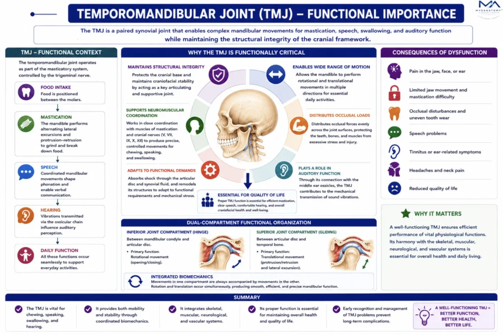
Functional Significance
Synovial joints provide the limited but essential mobility of the craniofacial skeleton. Through the temporomandibular joint and auditory ossicles, they enable mastication, speech, swallowing, auditory transmission, and neuromuscular coordination while preserving cranial stability and structural integrity.
OVERVIEW
Although synovial joints are relatively rare within the cranial skeleton, they perform critical physiological roles that enable dynamic functions of the head.
The skull is primarily designed as a rigid protective structure for the brain; however, certain functions – particularly mastication, speech articulation, and auditory transmission -require carefully controlled mobility. Synovial joints provide this mobility while maintaining the structural integrity of the cranial framework.
Within the skull, synovial joints are primarily associated with two functional systems:
the masticatory apparatus, represented by the temporomandibular joint (TMJ).
the auditory apparatus, represented by the synovial articulations between the middle ear ossicles.
Through these articulations, the skull integrates skeletal mechanics, neuromuscular coordination, and sensory processing, allowing complex physiological activities to occur within an otherwise rigid structure.
MASTICATORY INTEGRATION
Masticatory System
The temporomandibular joint operates as part of a neuromuscular masticatory system controlled by the trigeminal nerve.
Masseter – primary elevator of the mandible. Generates powerful bite force during chewing
Temporalis – elevation; retrusion. Posterior fibers stabilize the mandible during closure.
Medial Pterygoid – elevation-protrusion. Works with the masseter to form a muscular sling supporting the mandible.
Lateral Pterygoid – key muscle controlling disc and condyle movement; protrusion- depression; stabilization of articular disc. This muscle plays a major role in disc displacement disorders.
Exam Question
“Discuss the temporomandibular joint as an integrated neuromuscular component of the masticatory system, emphasizing the functional roles of the masseter, temporalis, medial pterygoid, and lateral pterygoid muscles in mandibular stabilization, force generation, disc control, and coordinated mastication.”
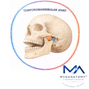
Masticatory Biomechanics
The TMJ operates in close coordination with dental occlusion.
During mastication:
food is positioned between the molars.
the mandible performs alternating lateral excursions.
one condyle rotates while the other translates.
teeth produce a grinding motion that breaks down food particles.
the TMJ therefore functions as part of a system involving:
teeth; mandible
cranial base; masticatory muscles
neuromuscular reflexes
Disruption of occlusion may alter joint biomechanics and produce temporomandibular disorders.
Exam Question
“Analyze the biomechanical relationship between the temporomandibular joint, dental occlusion, mandibular movement, and neuromuscular coordination during mastication, including the functional significance of alternating condylar rotation–translation dynamics and their role in efficient food processing.”
Masticatory Apparatus
The temporomandibular joint is the principal synovial joint responsible for mandibular mobility. It allows the mandible to move relative to the cranial base, enabling the mechanical processes required for food processing and verbal communication.
Through coordinated action of the muscles of mastication – including the masseter, temporalis, medial pterygoid, and lateral pterygoid – the TMJ enables a range of mandibular movements, including:
elevation and depression of the mandible
protrusion and retrusion
lateral excursion during chewing
These movements allow the mandible to generate the powerful occlusal forces required for mastication, while also permitting the fine adjustments needed for speech articulation.
During chewing, the mandible performs alternating lateral excursions, producing grinding movements of the molar teeth that break down food particles. This process is coordinated by reflex pathways within the trigeminal nerve (cranial nerve V), which regulates the activity of the masticatory muscles and modulates bite force according to the mechanical properties of the food being chewed.
In addition to mastication, the TMJ contributes to phonation and speech production. Precise mandibular positioning is necessary for articulation of many consonant sounds, particularly those involving the lips and teeth. Thus, the temporomandibular joint plays a fundamental role in human communication.
Furthermore, controlled mandibular depression and elevation are essential for swallowing (deglutition). Opening of the mouth permits food intake, while coordinated closure of the mandible contributes to the formation of the food bolus and initiation of swallowing.
Exam Question
“Explain how the temporomandibular joint functions as an integrated neuromuscular component of the masticatory apparatus, and analyze the coordinated biomechanical roles of the masseter, temporalis, medial pterygoid, and lateral pterygoid muscles in mandibular elevation, depression, protrusion, retrusion, and lateral excursion during mastication, speech, and swallowing.”
Auditory Apparatus
The synovial joints of the middle ear ossicles -specifically the incudomalleolar joint and the incudostapedial joint –play a crucial role in the mechanical transmission of sound.
The auditory ossicles form a chain linking the tympanic membrane to the oval window of the inner ear. Vibrations generated by sound waves striking the tympanic membrane are transmitted through this ossicular chain via small but precise movements at the synovial articulations.
The presence of synovial joints between the ossicles allows subtle adjustments in the relative positions of the malleus, incus, and stapes. These movements enable efficient transfer of vibrational energy and contribute to the mechanical amplification of sound waves before they reach the cochlea.
This mechanism compensates for the impedance mismatch between air vibrations in the external ear and fluid vibrations within the inner ear, thereby facilitating effective auditory perception.
Exam Question
“Discuss the biomechanical significance of the synovial articulations of the middle ear ossicles, and explain how movement at the incudomalleolar and incudostapedial joints contributes to mechanical amplification, impedance matching, and efficient transmission of vibrational energy from the tympanic membrane to the cochlea.”
ANATOMICAL RELATION
Anatomical Relation
Because of its location at the junction of the cranial base, infratemporal fossa, and parotid region, the TMJ is surrounded by several critical anatomical structures.
Anterior Relations
lateral pterygoid muscle– to the articular disc and condylar neck, allowing muscular control of disc movement.
infratemporal fossa
branches of the maxillary artery
Posterior Relations
external acoustic meatus
tympanic plate of temporal bone
auriculotemporal nerve
superficial temporal vessels
Because of these relationships, TMJ pathology may produce symptoms resembling ear disorders, including ear pain and tinnitus.
Medial Relations
maxillary artery – the maxillary artery may pass either medial or lateral to the mandibular neck, creating variation important in surgical approaches.
sphenomandibular ligament
Lateral Relations
parotid gland
facial nerve branches – passes through the parotid gland, making it vulnerable during TMJ surgery.
Exam Question
“Describe the anatomical relations of the temporomandibular joint within the infratemporal and parotid regions, and explain the clinical significance of its relationships to the lateral pterygoid muscle, maxillary artery, auriculotemporal nerve, parotid gland, facial nerve branches, and external acoustic meatus in temporomandibular disorders and surgical approaches.”
Neurovascular Integration
Innervation
the TMJ receives sensory innervation from branches of the mandibular division of the trigeminal nerve (CN V₃).
Major nerves include:
auriculotemporal nerve
masseteric nerve
deep temporal nerves
These nerves provide:
pain sensation; proprioception
reflex control of mandibular movement
This sensory network participates in the masticatory reflex system, which regulates biting force.
Blood Supply
Arterial supply arises primarily from branches of the:
maxillary artery
superficial temporal artery
Important branches include:
deep auricular artery
anterior tympanic artery
masseteric artery
Venous drainage
through veins that empty into the pterygoid venous plexus.
Exam Question
“Analyze the neurovascular organization of the temporomandibular joint, including its sensory innervation from branches of the mandibular division of the trigeminal nerve and arterial supply from branches of the maxillary and superficial temporal arteries, and discuss how this neurovascular integration contributes to proprioception, reflex control of mastication, pain transmission, and functional joint stability.”
SUMMARY TABLE