Conchae & Meatuses
The lateral wall of the nasal cavity gives rise to three curved bony projections, the superior, middle, and inferior nasal conchae, which divide the cavity into corresponding passageways known as the meatuses. These structures are not merely passive shelves; they are fundamental regulators of nasal airflow, surface area expansion, and mucosal conditioning. By producing turbulence, the conchae slow inspired air, increase contact between air and respiratory mucosa, and thereby optimize warming, humidification, and filtration before air reaches the lower respiratory tract
CORE


Overview
Anatomically, the superior and middle conchae belong to the ethmoid bone, whereas the inferior concha is an independent bone.
Their relationship to the meatuses is of major clinical importance because these channels serve as the principal routes for drainage of the paranasal sinuses and lacrimal apparatus.
ANATOMY
Superior Meatus
The superior meatus lies inferior to the superior nasal concha and forms a relatively small but clinically important recess within the upper part of the nasal cavity. It primarily receives drainage from the posterior ethmoidal air cells, linking the posterior ethmoid region to the nasal airway.
Because of its posterior and superior location, it is less accessible clinically than the middle meatus, yet remains important in understanding patterns of sinus disease.
Pathology involving the posterior ethmoidal sinuses may therefore present in relation to the superior meatus, especially in advanced inflammatory or obstructive states.
Exam Question
Explain the anatomical position of the superior meatus in relation to the superior nasal concha and analyze its role in the drainage of posterior ethmoidal air cells, including its clinical relevance in posterior sinus pathology.
Middle Meatus
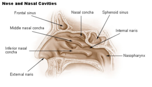
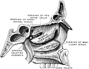
The middle meatus is the most functionally and clinically significant meatal compartment. Situated beneath the middle nasal concha, it contains the ostiomeatal complex, a highly organized drainage corridor through which several major sinuses communicate with the nasal cavity.
It receives drainage from the frontal sinus, maxillary sinus, and anterior ethmoidal air cells; in broader anatomical context, the middle meatus is also closely related to the middle ethmoidal cells.
This region therefore represents the principal drainage hub of the paranasal sinus system. Because ventilation and mucus clearance of multiple sinuses depend on its patency, even minor mucosal edema or anatomical narrowing within the middle meatus may predispose to sinus obstruction, secretion retention, and chronic rhinosinusitis. For this reason, the middle meatus is central in endoscopic anatomy and sinus surgery.
Exam Question
Describe the anatomical organization of the middle meatus and explain why it functions as the principal drainage hub of the paranasal sinuses, including its relationship to the ostiomeatal complex.
Inferior Meatus
The inferior meatus lies beneath the inferior nasal concha and is structurally simpler than the middle meatus, but it remains of major physiological importance.
It receives the terminal opening of the nasolacrimal duct, which drains tears from the lacrimal sac into the nasal cavity.
This anatomical relationship explains the intimate connection between lacrimal and nasal physiology: excess tears may enter the inferior meatus, and nasal mucosal disease may secondarily influence lacrimal drainage.
Obstruction of this passage contributes to tear overflow and recurrent lacrimal stagnation.
Exam Question
Describe the anatomical features of the inferior meatus and explain the functional and clinical significance of the nasolacrimal duct opening within it, particularly in relation to tear drainage and epiphora.
OPENINGS & COMMUICATIONS
General
The nasal cavity is not an isolated chamber; rather, it functions as a central crossroads between the external environment, paranasal sinuses, nasopharynx, orbit, and cranial base.
Its openings and communications allow airflow conduction, mucus drainage, olfactory transmission, and tear passage.
From a regional anatomical perspective, the nasal cavity must therefore be understood as both a respiratory passage and a communication corridor linking multiple craniofacial compartments. These communications also explain why disease processes of the nose may extend to adjacent spaces, including the sinuses and orbit.
Exam Question
Explain how the nasal cavity functions as a central anatomical crossroads, detailing how its openings and communications enable airflow conduction, mucus drainage, olfactory transmission, and tear passage.
Choanae
The choanae are the paired posterior apertures of the nasal cavity that establish continuity with the nasopharynx. They form the posterior limit of the nasal chambers and are essential for routing inspired and expired air between the nasal passages and the pharyngeal airway.
Anatomically, they are bounded superiorly by the sphenoid, inferiorly by the palatine bones, laterally by the medial pterygoid plates, and medially by the vomer.
These boundaries reflect the integration of the nasal cavity with the posterior facial skeleton and skull base. Functionally, the choanae are crucial for nasal breathing, while clinically they are important in congenital obstruction, posterior nasal masses, and endoscopic orientation.
Exam Question
Describe the anatomical boundaries of the choanae and explain their functional role in establishing continuity between the nasal cavity and nasopharynx, including their importance in airflow routing.
Paranasal Sinuses
The paranasal sinuses drain into the nasal cavity through highly specific ostia that empty into different meatal compartments. These drainage pathways are essential for sinus aeration, mucus clearance, and pressure equilibration. The anatomical arrangement is not random; it reflects both embryological development and the functional architecture of sinonasal ventilation.
Drainage pathways
Frontal sinus → middle meatus
Maxillary sinus → middle meatus
Anterior ethmoidal sinuses → middle meatus
Posterior ethmoidal sinuses → superior meatus
Exam Question
Explain the organization of paranasal sinus drainage into the nasal cavity, detailing how specific sinuses relate to different meatal compartments and how this arrangement supports ventilation and mucus clearance.
Sphenoidal Sinuses
This organization makes the middle meatus the dominant drainage zone of the paranasal sinus system.
The sphenoethmoidal recess, located superior and posterior to the superior concha, specifically receives the sphenoidal sinus and is therefore especially relevant in skull base and transsphenoidal anatomy.
Exam Question
Describe the location of the sphenoethmoidal recess and explain its role in sphenoidal sinus drainage, including its anatomical and clinical relevance.
Nasolacrimal Canal
The nasolacrimal duct drains tears from the lacrimal sac into the inferior nasal meatus, establishing an important physiological link between the orbit and nasal cavity.
This pathway allows tears to pass from the ocular surface into the nasal passage, where they contribute to mucosal moisture and are ultimately cleared.
The anatomical continuity of the lacrimal system with the inferior meatus explains why crying is accompanied by increased nasal discharge and why obstruction of the duct leads to epiphora.
From a clinical standpoint, the nasolacrimal canal is important in lacrimal drainage disorders, recurrent dacryocystitis, and procedures involving the inferior meatus.
Exam Question
Describe the anatomical pathway of the nasolacrimal duct and explain its functional connection between the lacrimal system and the nasal cavity, including its clinical significance in tear drainage disorders.
SUMMARY TABLE
