
Plexus Organization
Nerve plexuses are networks formed by the anterior (ventral) rami of spinal nerves, in which axons from multiple spinal segments converge, intermingle, and redistribute to form peripheral nerves carrying motor, sensory, and autonomic fibersto the limbs and anterior trunk.
Functionally, they provide multisegmental innervation to muscles, joints, and skin, enabling precise motor control, integrated proprioception, and coordinated reflex activity across functionally related structures.
A defining feature is fiber redistribution, whereby each spinal nerve contributes to multiple peripheral nerves, and each peripheral nerve contains fibers from several spinal segments, ensuring functional redundancy and resilience against single-segment injury.
In contrast to segmental posterior rami, anterior rami form the major plexuses – cervical (C1-C4), brachial (C5-T1), lumbar (L1–L4), and sacral (L4-S4) – which supply regions requiring complex neuromechanical coordination.
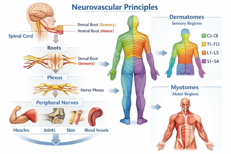
AI-generated illustration ( MyoAnatomy)
Hierarchical Organization
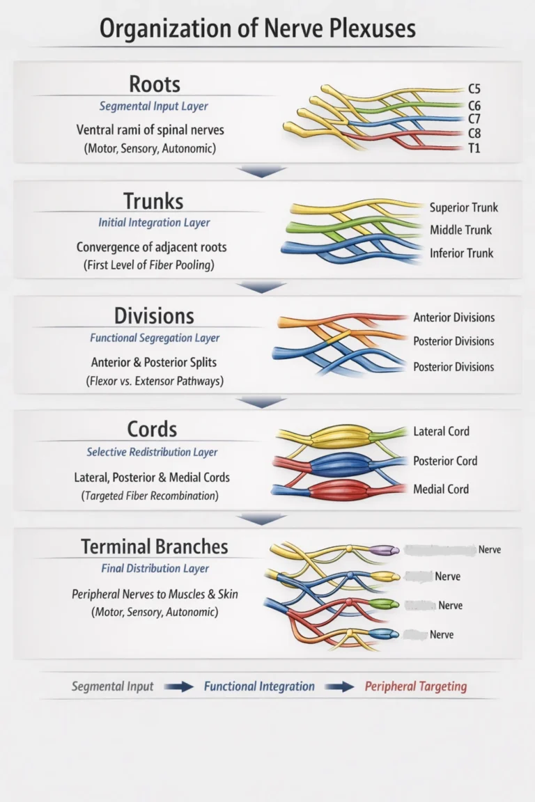
Nerve plexuses constitute hierarchically organized redistribution networks of the anterior (ventral) rami, arranged into sequential levels—roots → trunks → divisions → cords → terminal branches—that enable progressive fiber convergence, recombination, functional segregation, and precise peripheral targeting. This architecture transforms segmentally organized spinal nerve output into a regionally integrated neural system adapted to the structural and functional demands of the limbs.
At each level of the plexus, mixed neural components – somatic motor (efferent), somatic sensory (afferent), and autonomic (primarily sympathetic) fibers – undergo controlled reorganization. Initially, roots preserve segmental identity, but subsequent convergence within trunks and redistribution through divisions and cords allows selective sorting of fibers according to functional role and anatomical destination.
A critical organizational principle is the segregation into anterior and posterior divisions, which reflects embryological patterning and aligns with biomechanical function: anterior divisions predominantly supply flexor compartments, whereas posterior divisions supply extensor compartments. This ensures that neural output is matched to coordinated movement patterns and compartment-based muscle function.
Through this multilevel redistribution, each terminal peripheral nerve contains a precisely configured mixture of fibers derived from multiple spinal segments, enabling multisegmental innervation, coordinated activation of muscle groups across joints, and integrated sensory feedback.
Thus, nerve plexuses represent a highly efficient neuroanatomical system that converts segmental neural signals intofunctionally specialized, biomechanically aligned, and clinically resilient pathways, forming the foundation for coordinated movement, proprioception, and adaptive neuromuscular control
AI-generated illustration ( MyoAnatomy)
Roots
Segmental Input Layer
Roots represent the anterior (ventral) rami of spinal nerves, carrying mixed somatic motor (α-motor), somatic sensory (afferent), and autonomic (sympathetic) fibers from individual spinal segments. They preserve the segmental origin of neural output, serving as the primary input streams that feed into the plexus. At this level, fibers are still organized according to their spinal level, providing the anatomical basis for dermatomes and myotomes.
Trunks
Initial Integration Layer
Trunks are formed by the convergence of adjacent roots, creating composite neural pathways that integrate input from multiple spinal segments. This stage represents the first level of functional pooling, allowing early combination of fibers destined for related anatomical regions. Trunks reduce strict segmental separation and initiate the transition toward region-based neural distribution.
Devisions
Functional Segregation Layer
Each trunk divides into anterior and posterior divisions, representing a critical step of functional separation based on embryological and biomechanical organization.
Anterior divisions → supply flexor (ventral) compartments
Posterior divisions → supply extensor (dorsal) compartments
This division aligns neural output with movement patterns and compartmental function, organizing fibers according to their biomechanical role.
Cords
Selective Redistribution layer
Divisions reorganize into cords, named relative to a central artery (e.g., axillary artery). At this level, fibers undergo selective regrouping based on both function and target distribution, producing pathways with specific combinations of segmental inputs. Cords represent a higher-order integration stage where neural elements are arranged to supply coordinated muscle groups and joint systems.
Terminal Branches
Terminal branches are the peripheral nerves that arise from the cords, containing precisely arranged mixtures of fibers from multiple spinal segments. They deliver motor, sensory, and autonomic innervation to defined muscular, articular, and cutaneous territories. This final stage reflects complete functional specialization, enabling coordinated movement, precise sensory feedback, and efficient neuromuscular control.