Brachial Plexus
MYO CORE
Clinical Significance
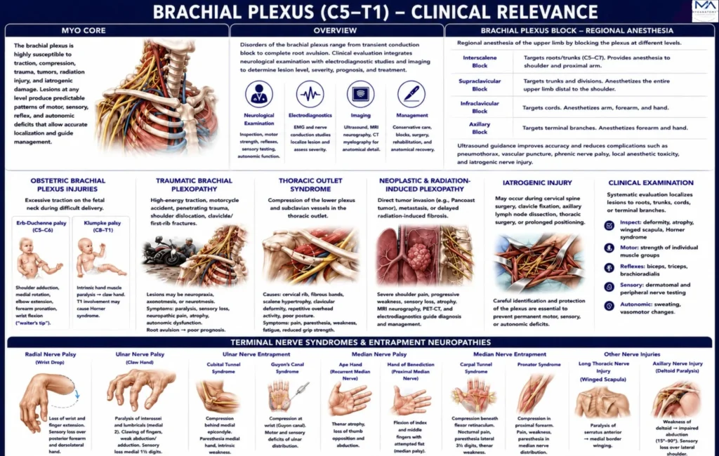
The brachial plexus is one of the most clinically significant neural networks of the peripheral nervous system because its long anatomical course from the cervical spine to the axilla exposes it to traction, compression, penetrating trauma, neoplastic infiltration, radiation injury, and iatrogenic damage. Lesions may occur at the level of the roots, trunks, divisions, cords, or terminal branches, each producing characteristic patterns of motor weakness, sensory impairment, reflex abnormalities, and autonomic dysfunction.
OVERVIEW
Clinical disorders of the brachial plexus range from transient conduction block to complete spinal root avulsion.
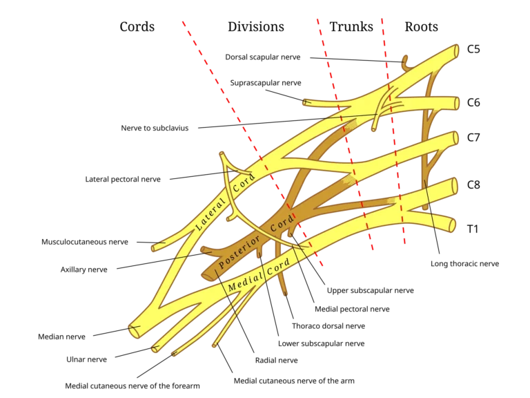
Because every terminal peripheral nerve contains fibers originating from multiple spinal cord segments, injury produces predictable combinations of muscular paralysis, sensory deficits, and loss of coordinated upper-limb function rather than isolated impairment of a single muscle.
Modern diagnosis integrates neurological examination with electromyography, nerve conduction studies, ultrasonography, CT myelography, and magnetic resonance neurography to determine lesion level, evaluate severity, estimate prognosis, and guide conservative or surgical management.
Source: Gray’s Anatomy (1918), illustration by Henry Vandyke Carter. Vector adaptation by Mattopaedia and MissMJ via Wikimedia Commons. Public Domain.
Exam Question
How does the multisegmental organization of the brachial plexus influence the pattern of motor and sensory deficits following injury, and why are neurological examination, electrodiagnostic studies, and advanced imaging essential for accurate lesion localization, prognostic assessment, and clinical management?
ANATOMY
Obstetric Plexopathy
Obstetric brachial plexus injury usually results from traction on the fetal neck or shoulder during difficult delivery, particularly shoulder dystocia. Injury may range from neurapraxia to root rupture or avulsion.
Erb-Duchenne palsy (C5–C6) weakens shoulder abduction and lateral rotation, elbow flexion, and forearm supination, producing the classic waiter’s-tip posture. Klumpke palsy (C8-Th1) predominantly affects the intrinsic hand muscles, causing clawing; associated Th1 sympathetic injury may produce Horner syndrome.
Early neurological assessment and rehabilitation are essential, with microsurgical reconstruction considered for severe nonrecovering lesions.
Exam Question
How does the segmental anatomy of the upper and lower brachial plexus explain the contrasting motor deficits of Erb–Duchenne and Klumpke palsies, and why may T1 injury produce Horner syndrome?
Traumatic Brachial Plexopathy
Traumatic brachial plexopathy follows high-energy traction, motorcycle collision, penetrating injury, shoulder dislocation, or clavicular and first-rib fractures.
Lesions range from transient conduction block to rupture or root avulsion and may produce flaccid weakness, sensory loss, reflex depression, neuropathic pain, muscle atrophy, and autonomic disturbance.
Preganglionic root avulsion has a poorer prognosis than postganglionic injury because continuity with the spinal cord is lost and direct regeneration across the avulsed root is not possible. MRI neurography, CT myelography, electromyography, and nerve-conduction studies define lesion level and guide nerve grafting, transfer, or reconstructive procedures.
Exam Question
How do mechanism, lesion level, and preganglionic versus postganglionic anatomy determine prognosis and reconstructive strategy in traumatic brachial plexopathy?
Thoracic Outlet Syndrome
Thoracic outlet syndrome results from compression of the lower plexus, often with the subclavian vessels, within the interscalene triangle, costoclavicular interval, or retropectoralis minor space.
Cervical ribs, fibrous bands, scalene hypertrophy, clavicular deformity, repetitive overhead activity, and postural dysfunction may reduce these spaces.
Neurogenic compression produces pain, paresthesia, intrinsic hand weakness, fatigue, and reduced grip strength, whereas vascular involvement may cause ischemia, swelling, or venous congestion.
Management begins with postural correction and physiotherapy; progressive neurological or vascular compromise may require decompression.
Exam Question
Why is the lower brachial plexus particularly vulnerable within the thoracic outlet, and how do its relationships with the subclavian vessels explain combined neurological and vascular symptoms?
Brachial Plexus Block
Brachial plexus blockade provides regional anesthesia for upper-limb procedures by targeting the plexus at different anatomical levels.
Interscalene, supraclavicular, infraclavicular, and axillary approaches produce distinct anesthetic territories because they engage roots or trunks, compact plexus elements, cords, or terminal branches, respectively.
Ultrasound guidance improves precision and reduces complications, including vascular puncture, pneumothorax, phrenic nerve paresis, local-anesthetic systemic toxicity, and direct neural injury.
Exam Question
How does the anatomical level of each brachial plexus block determine anesthetic distribution, incomplete blockade patterns, and procedure-specific complications?
Neoplastic Plexopathy
The brachial plexus may be affected by direct tumor invasion, metastatic disease, nerve-sheath tumors, operative injury, or delayed radiation fibrosis.
Pancoast tumors at the lung apex commonly involve the lower trunk and sympathetic chain, producing severe shoulder pain, C8-Th1 sensory and motor deficits, intrinsic hand weakness, and Horner syndrome.
Neoplastic plexopathy is often painful and progressive, whereas radiation-induced plexopathy more commonly evolves gradually through fibrosis, microvascular injury, and ischemia.
MRI neurography, PET-CT, and electrodiagnostic studies assist differentiation and guide oncological or surgical management
Exam Question
How do the anatomical relationships of the lower trunk and sympathetic chain explain the presentation of Pancoast tumors, and which clinical and imaging features help distinguish neoplastic from radiation-induced plexopathy?
Clinical Examination
Clinical examination integrates inspection, muscle testing, reflexes, dermatomal and peripheral sensory assessment, and autonomic evaluation to distinguish radiculopathy, plexopathy, and isolated terminal-nerve lesions.
Radial nerve palsy causes wrist drop through paralysis of the wrist and finger extensors.
Median nerve injury may produce thenar atrophy, loss of opposition, ape-hand deformity, or the hand of benediction; distal compression causes carpal tunnel syndrome, while proximal compression may produce pronator syndrome.
Ulnar nerve palsy causes intrinsic hand weakness and clawing, while entrapment may occur in the cubital tunnel or Guyon canal.
Long thoracic nerve injury produces scapular winging through serratus anterior paralysis. These characteristic patterns permit anatomical localization and guide electrodiagnostic testing, imaging, rehabilitation, and surgery
Exam Question
How do wrist drop, ape hand, hand of benediction, claw hand, winged scapula, and carpal tunnel syndrome localize injury to specific terminal branches and distinguish distal neuropathy from proximal brachial plexus disease?
SUMMARY TABLE