Brachial Plexus
MYO CORE
Branches
The brachial plexus gives rise to an organized network of collateral and terminal branches that redistribute fibers from the anterior (ventral) rami of C5 – Th1, establishing the principal peripheral nerves of the upper limb and providing integrated motor, sensory, and sympathetic innervation throughout the shoulder girdle and upper extremity.
OVERVIEW
The branches of the brachial plexus represent the definitive peripheral redistribution of segmental spinal nerve fibers following hierarchical organization of the plexus.
Arising sequentially from the roots, trunks, cords, and terminal divisions, they establish a highly integrated neural network that coordinates voluntary movement, cutaneous sensation, proprioception, articular innervation, and autonomic regulation throughout the upper limb.
Proximal collateral branches primarily stabilize the scapulothoracic articulation and shoulder girdle, whereas terminal branches provide the principal neurovascular supply to the arm, forearm, and hand.
This precise anatomical organization permits efficient biomechanical coordination, preserves neurological function through intersegmental fiber integration, and provides the structural basis for lesion localization, peripheral nerve reconstruction, regional anesthesia, and advanced neuroimaging.
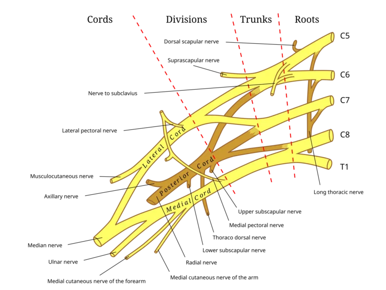
Source: Gray’s Anatomy (1918), illustration by Henry Vandyke Carter. Vector adaptation by Mattopaedia and MissMJ via Wikimedia Commons. Public Domain.
Exam Question
How does the hierarchical branching organization of the brachial plexus optimize intersegmental fiber integration, upper-limb biomechanics, and anatomical localization of peripheral nerve lesions?
ANATOMY
Branch Classification
The branches of the brachial plexus areanatomically classified into supraclavicular collateral, infraclavicular collateral, and terminal branches according to their level of origin, topographical course, and peripheral distribution.
This hierarchical classification reflects the progressive reorganization of segmental spinal nerve fibers into functionally specialized peripheral nerves while maintaining orderly anatomical relationships throughout the cervical and axillary regions.
Exam Question
How does the anatomical classification of brachial plexus branches reflect their developmental organization, peripheral distribution, and functional specialization?
Supraclavicular Branches
The supraclavicular collateral branches arise from the roots and upper trunk before the plexus enters the cervicoaxillary canal. These include the dorsal scapular, long thoracic, suprascapular, and nerve to subclavius, together with direct muscular branches.
They establish the neuromuscular framework responsible for scapulothoracic stability, glenohumeral biomechanics, and coordinated scapulohumeral rhythm, while providing motor innervation to the deep cervical, scapular, and proximal thoracic musculature.
Because they originate proximal to the clavicle, isolated injury commonly indicates root or upper trunk pathology rather than distal peripheral nerve lesions.
Exam Question
How do the supraclavicular collateral branches contribute to scapulothoracic stability and shoulder biomechanics, and why do their injury patterns facilitate localization of proximal brachial plexus lesions?
Infraclavicullar Branches
The infraclavicular collateral branches originate from the lateral, posterior, and medial cords within the axillary sheath surrounding the second part of the axillary artery.
They include the lateral and medial pectoral, upper and lower subscapular, thoracodorsal, medial brachial cutaneous, and medial antebrachial cutaneous nerves. These branches innervate the pectoral girdle, posterior axillary musculature, and medial cutaneous territories of the arm and forearm while maintaining close anatomical relationships with the axillary vessels.
Their topographical position makes them particularly susceptible to injury during axillary trauma, lymph node dissection, breast surgery, and vascular procedures.
Exam Question
How do the anatomical relationships of the infraclavicular collateral branches influence surgical exposure, regional anesthesia, and localization of axillary nerve injuries?
Terminal Branches
The terminal branches represent the final peripheral projection of the brachial plexus and comprise the musculocutaneous, axillary, median, radial, and ulnar nerves.
Each nerve contains fibers derived from multiple spinal cord segments, allowing coordinated innervation of the shoulder, arm, forearm, and hand while preserving functional redundancy through intersegmental integration.
Their mixed composition explains why spinal root lesions and peripheral nerve injuries produce distinct patterns of motor and sensory impairment.
Exam Question
How does the multisegmental composition of the terminal branches explain the characteristic differences between spinal root lesions and isolated peripheral nerve injuries?
Functional Organization
The branches of the brachial plexus function as integrated mixed peripheral nerves that coordinate voluntary movement, cutaneous sensation, proprioceptive feedback, articular innervation, and sympathetic regulation throughout the upper extremity.
This organization synchronizes shoulder stability, upper-limb biomechanics, fine motor control, and sensory perception while maintaining vascular homeostasis and integumentary function through accompanying sympathetic fibers.
Exam Question
How does the functional organization of the brachial plexus coordinate movement, proprioception, sensory integration, and sympathetic regulation to optimize upper-limb performance?
Anatomical Variations
The branching pattern of the brachial plexus demonstrates considerable anatomical variability involving root contributions, trunk formation, cord configuration, collateral branching, and communications between terminal nerves.
Common variants include prefixed and postfixed plexuses, musculocutaneous–median nerve communications, and clinically important anastomoses such as the Martin-Gruber, Marinacci, Riche-Cannieu, and Berrettini communications.
Recognition of these variations is fundamental for peripheral nerve surgery, microsurgical reconstruction, electrodiagnostic interpretation, regional anesthesia, and high-resolution neuroimaging, where atypical anatomy may substantially influence both diagnosis and operative planning.
Exam Question
Which anatomical variations of the brachial plexus have the greatest influence on peripheral nerve surgery, electrodiagnostic interpretation, regional anesthesia, and advanced neuroimaging, and why?
SUMMARY TABLE