Internal Carotid Artery
MYO CORE
Branches & Distribution
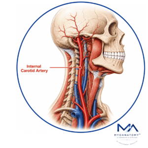
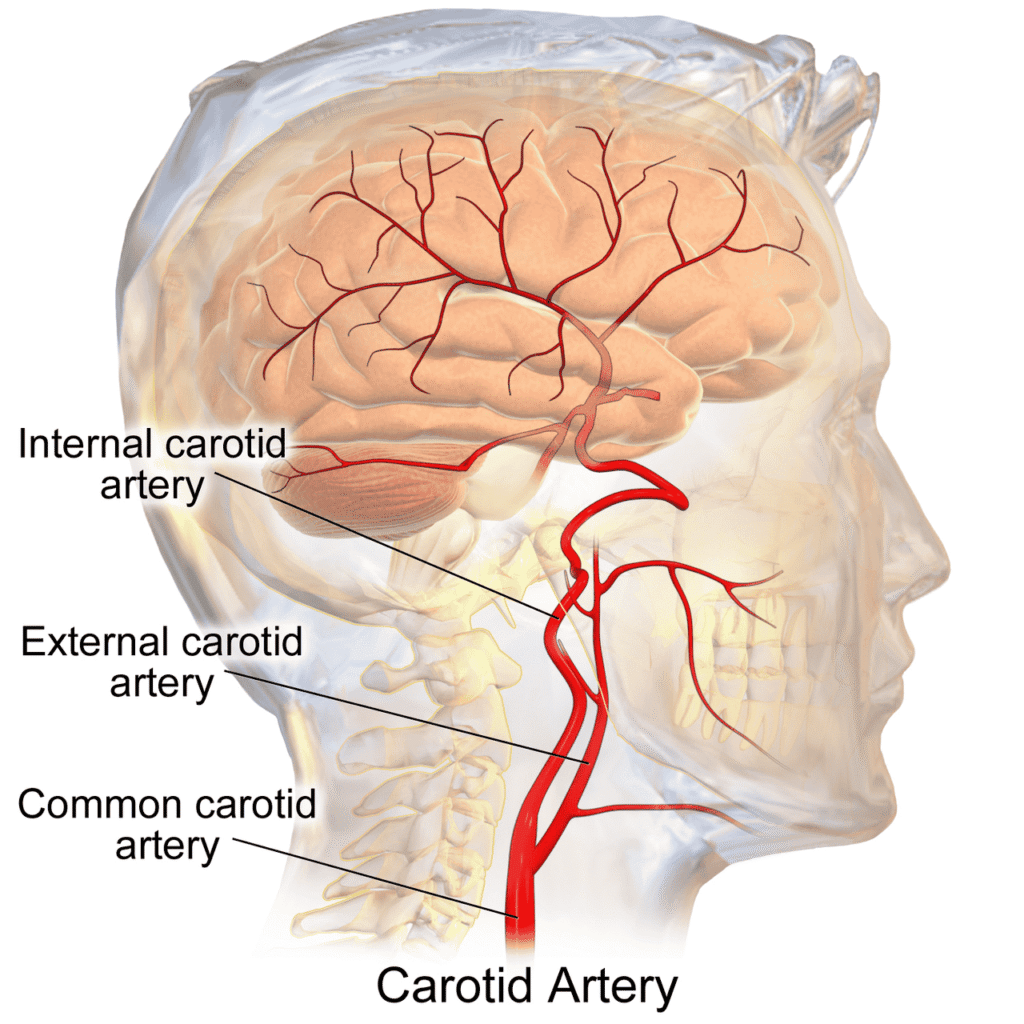
The internal carotid artery undergoes a specialized transition from a major extracranial conduit vessel to a complex intracranial distribution network. Through its sequential branches, it supplies the orbit, visual apparatus, deep cerebral structures, and cerebral cortex, thereby supporting vision, neuroendocrine regulation, higher cognition, sensory integration, and voluntary motor function.
OVERVIEW
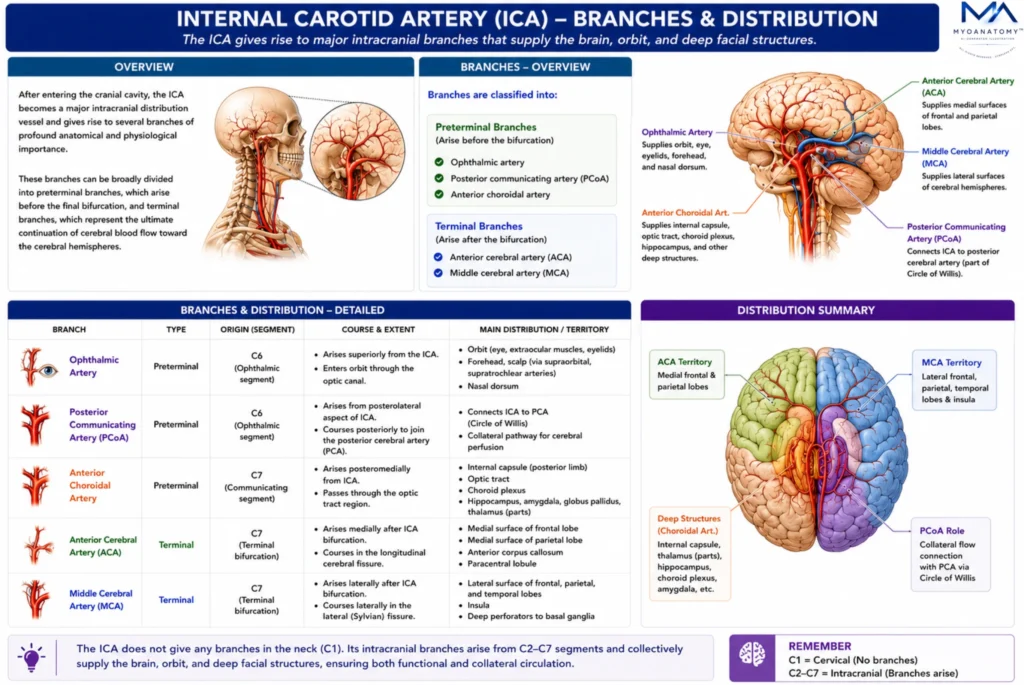
Following its entrance into the cranial cavity, the internal carotid artery becomes a major intracranial distribution vessel and gives rise to several branches of profound anatomical and physiological importance.
These branches can be broadly divided into preterminal branches, which arise before the final bifurcation, and terminal branches, which represent the ultimate continuation of cerebral blood flow toward the cerebral hemispheres.
The principal preterminal branches include the ophthalmic artery, posterior communicating artery, and anterior choroidal artery. Together these vessels supply the orbit, retina, visual pathways, hypothalamus, ventricular system, internal capsule, basal ganglia, and multiple deep cerebral structures.
The ICA ultimately terminates by dividing into the anterior cerebral artery and middle cerebral artery, which collectively supply the majority of the cerebral cortex.
This branching pattern creates an integrated vascular network that links the orbit, diencephalon, deep gray matter, and cerebral hemispheres into a unified circulatory system capable of supporting the highly specialized functions of the human brain.
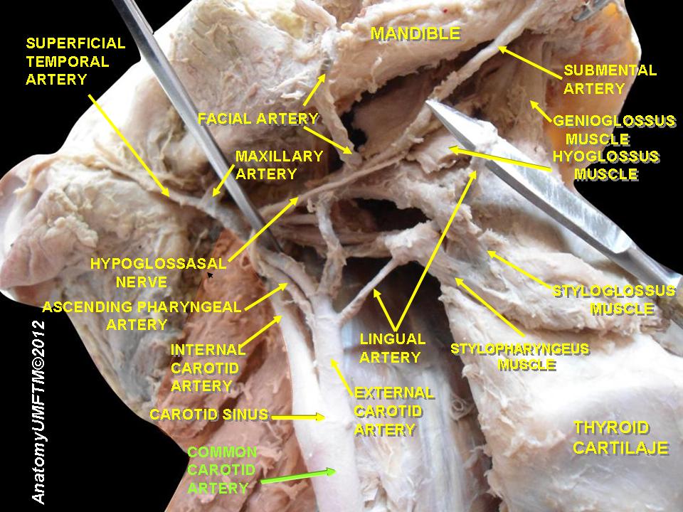
Internal Carotid Branches
Ophthalmic Artery
The ophthalmic artery is typically the first major intracranial branch of the internal carotid artery, arising from the supraclinoid segment immediately after the ICA enters the subarachnoid space.
It accompanies the optic nerve through the optic canal and becomes the principal arterial supply of the orbit and visual apparatus. Through its numerous branches, it vascularizes the retina, optic nerve, extraocular muscles, lacrimal gland, eyelids, conjunctiva, orbital connective tissues, forehead, and dorsum of the nose.
Functionally, the ophthalmic artery represents the vascular gateway of vision. Its central retinal branch provides the sole arterial supply to the inner retina, a tissue characterized by exceptionally high metabolic demand and minimal collateral circulation.
Consequently, interruption of ophthalmic or retinal blood flow may rapidly result in irreversible monocular visual loss. Extensive anastomoses with branches of the external carotid artery additionally create important collateral pathways between intracranial and extracranial circulations, particularly during proximal internal carotid artery stenosis.
Exam Question
Analyze the ophthalmic artery as the principal vascular gateway to the orbit and retina, explaining its anatomical course, major territories supplied, collateral significance, and the visual consequences of proximal or distal occlusion.
Posterior Communicating Artery
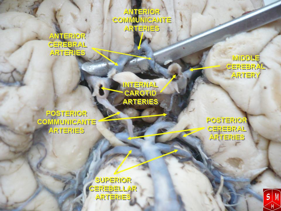
The posterior communicating artery (PCoA) arises from the posteromedial wall of the internal carotid artery and courses posteriorly to join the posterior cerebral artery.
Although usually small in caliber, it constitutes one of the most important collateral vessels in the cerebral circulation by linking the carotid and vertebrobasilar systems within the Circle of Willis.
Beyond its collateral role, the PCoA gives rise to perforating arteries supplying the optic tract, hypothalamus, mammillary bodies, thalamus, and adjacent diencephalic structures.
These territories participate in autonomic regulation, endocrine control, memory processing, and visual integration.
Clinically, the ICA-PCoA junction is a common site of saccular aneurysm formation; aneurysmal expansion may compress the adjacent oculomotor nerve, producing ipsilateral ptosis, ophthalmoplegia, and a dilated non-reactive pupil.
Exam Question
Assess the anatomical and physiological importance of the posterior communicating artery within the Circle of Willis, and explain how aneurysmal pathology at the ICA–PCoA junction may produce characteristic neuro-ophthalmological deficits.
Anterior Choroidal Artery
The anterior choroidal artery is a small yet disproportionately important branch arising distal to the posterior communicating artery. It courses posterolaterally along the optic tract toward the temporal horn of the lateral ventricle, supplying a highly concentrated collection of critical neuroanatomical structures.
Its vascular territory includes the optic tract, lateral geniculate body, posterior limb of the internal capsule, globus pallidus, cerebral peduncle, hippocampus, amygdala, portions of the thalamus, and the choroid plexus of the lateral ventricle.
Because the posterior limb of the internal capsule contains densely packed corticospinal, corticobulbar, and thalamocortical fibers, even small infarctions may produce severe neurological deficits.
Occlusion classically results in Anterior Choroidal Artery Syndrome, characterized by contralateral hemiplegia, hemisensory loss, and homonymous hemianopia due to simultaneous involvement of motor, sensory, and visual pathways.
Exam Question
Evaluate the disproportionate clinical importance of the anterior choroidal artery relative to its size, discussing its vascular territory, relationship to major motor and visual pathways, and the neurological syndrome produced by its occlusion.
Anterior Cerebral Artery
The anterior cerebral artery is one of the terminal branches of the internal carotid artery and serves as the primary arterial supply to the medial surfaces of the frontal and parietal lobes.
After arising from the ICA, it courses anteromedially above the optic chiasm and enters the longitudinal cerebral fissure, where it is connected to its contralateral counterpart by the anterior communicating artery.
The ACA supplies the cingulate gyrus, corpus callosum, medial frontal cortex, medial parietal cortex, and paracentral lobule. These regions are essential for executive function, motivation, emotional behavior, lower-limb motor control, somatosensory integration, and voluntary bladder regulation.
Consequently, ACA infarction characteristically produces contralateral lower-limb weakness and sensory loss, often accompanied by abulia, personality change, impaired judgment, or urinary incontinence due to frontal lobe involvement.
Exam Question
Analyze the anterior cerebral artery as the principal arterial supply of the medial cerebral hemisphere, and explain how interruption of its circulation affects executive function, behavior, lower-limb motor control, sensation, and continence.
Middle Cerebral Artery
The middle cerebral artery is the larger terminal branch and direct continuation of the internal carotid artery. After entering the lateral sulcus, it divides into an extensive network that supplies the majority of the lateral cerebral hemisphere, making it the dominant distribution artery of the forebrain.
Its cortical branches vascularize much of the frontal, parietal, and temporal lobes, including the primary motor cortex, primary somatosensory cortex, frontal eye fields, Broca area, Wernicke area, auditory cortex, and multimodal association regions. Deep perforating lenticulostriate arteries additionally supply the basal ganglia and internal capsule.
Owing to its extensive vascular territory and the functional importance of the regions supplied, the MCA is the vessel most frequently involved in ischemic stroke.
Occlusion may produce contralateral hemiplegia, hemisensory loss, aphasia, hemispatial neglect, gaze deviation, and profound cognitive impairment depending on the hemisphere affected.
Exam Question
Discuss why the middle cerebral artery is considered the most clinically significant terminal branch of the internal carotid artery, correlating its extensive cortical and deep perforating territories with the neurological deficits observed following MCA infarction.
SUMMARY TABLE