External Carotid Artery
MYO CORE
Branches & Distribution
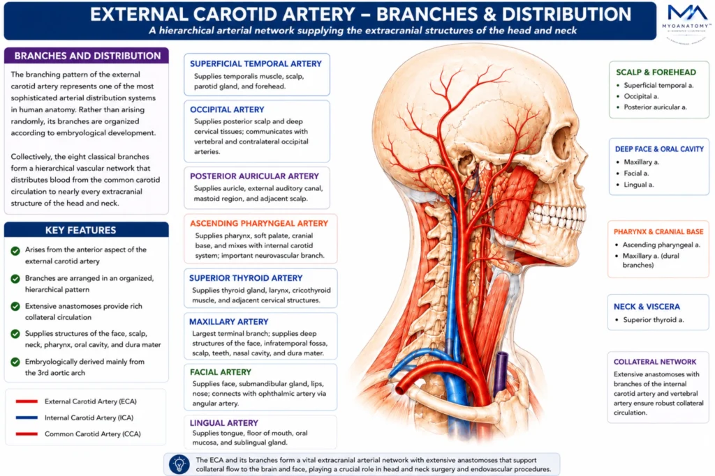
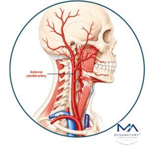
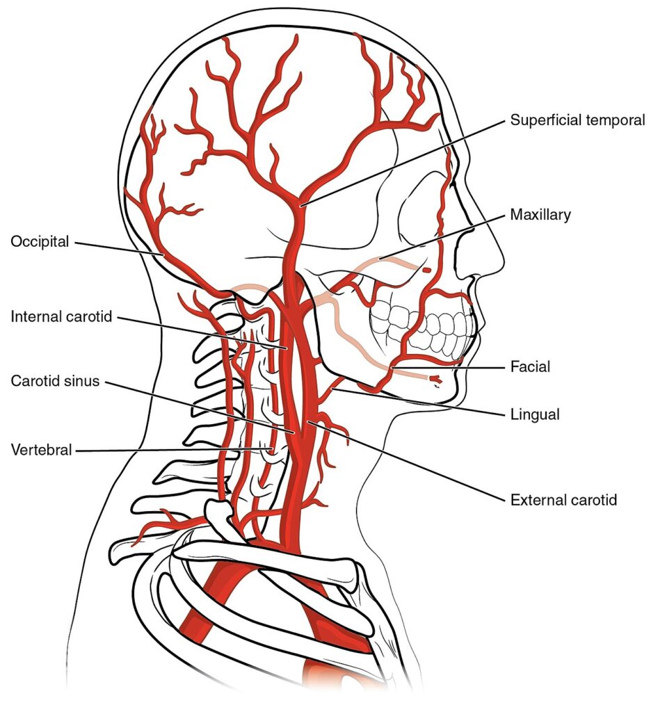
The external carotid artery exhibits a highly organized branching architecture, with its vessels distributed according to embryological and regional developmental patterns. Together, its eight classical branches form a hierarchical arterial network that supplies nearly all extracranial structures of the head and neck.
OVERVIEW
The branching pattern of the external carotid artery represents one of the most sophisticated arterial distribution systems in human anatomy.
Rather than arising randomly, its branches are organized according to embryological development, regional vascular demand, and anatomical orientation relative to the parent vessel.
Collectively, the 8 classical branches form a hierarchical vascular network that distributes blood from the common carotid circulation to nearly every extracranial structure of the head and neck.
This arrangement reflects the functional requirements of speech, mastication, swallowing, facial expression, sensory processing, thermoregulation, and endocrine activity.
Anterior Branches
Superior Thyroid Artery
The superior thyroid artery is typically the first branch of the external carotid artery and serves as the dominant arterial supply of the thyroid gland and adjacent cervical viscera.
As it descends toward the superior pole of the thyroid gland, it distributes branches to the larynx, upper trachea, infrahyoid musculature, sternocleidomastoid muscle, and cricothyroid region.
Through its superior laryngeal branch, the artery accompanies the internal laryngeal nerve through the thyrohyoid membrane, forming a clinically important neurovascular relationship encountered during thyroid and laryngeal surgery.
Functionally, the superior thyroid artery maintains the exceptionally high metabolic demands of thyroid tissue and contributes significantly to the vascular support of phonatory and respiratory structures.
Its extensive anastomoses with the inferior thyroid artery create an important collateral pathway within the endocrine vascular system of the neck.
Exam Question
Analyze how the anatomical course and branching pattern of the superior thyroid artery support the metabolic and functional demands of the thyroid gland, larynx, and adjacent cervical structures.
Lingual Artery
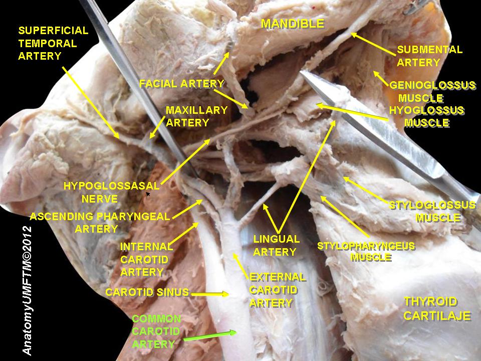
The lingual artery constitutes the principal arterial supply of the tongue and floor of the mouth. Originating at the level of the greater horn of the hyoid bone, it passes deep to the hypoglossus muscle before entering the substance of the tongue, where it divides into numerous terminal branches.
Its vascular territory includes the intrinsic and extrinsic tongue musculature, sublingual gland, oral mucosa, lingual gingiva, and floor of the oral cavity. Through its deep lingual branches, the artery supports one of the most metabolically active muscular organs in the human body.
This extensive vascularization is essential for the continuous muscular activity required for speech articulation, swallowing, mastication, and taste-related functions.
The lingual artery also forms part of a dense arterial network within the oral cavity, ensuring uninterrupted perfusion despite the considerable mechanical forces generated during chewing and tongue movement.
Exam Question
Explain how the deep anatomical course of the lingual artery enables efficient vascularization of the tongue while protecting the vessel during complex movements associated with speech, mastication, and swallowing.
Facial Artery
The facial artery is the largest anterior branch and the dominant arterial supply of the superficial face.
Characterized by its highly tortuous course, the vessel traverses the submandibular region before winding around the inferior border of the mandible and ascending toward the medial canthus of the eye.
Its distribution encompasses the lips, nose, cheeks, facial musculature, submandibular gland, tonsillar region, soft palate, and extensive areas of facial skin. The remarkable tortuosity of the vessel serves as an adaptive mechanism that accommodates continuous movement associated with mastication, speech, swallowing, and facial expression without compromising blood flow.
Through numerous anastomotic connections with branches of the ophthalmic artery, the facial artery forms a major communication between the external and internal carotid arterial systems, making it one of the most significant vessels involved in collateral craniofacial circulation.
Exam Question
Evaluate the anatomical significance of the facial artery’s tortuous course and discuss how its extensive anastomotic network contributes to facial perfusion and collateral circulation.
Posterior Branches
Occipital Artery
The occipital artery courses posteriorly toward the occipital region and serves as the principal arterial supply of the posterior scalp and deep cervical tissues.
During its course, it provides branches to the sternocleidomastoid muscle, splenius capitis, longissimus capitis, and numerous deep muscles of the posterior neck.
Beyond its regional perfusion role, the occipital artery participates extensively in collateral circulation through anastomoses with vertebral, deep cervical, and contralateral occipital arterial systems.
These communications become particularly important when major cervical arteries are compromised, allowing redistribution of blood flow between extracranial and vertebrobasilar territories
Exam Question
Discuss the role of the occipital artery in posterior scalp vascularization and explain its importance within extracranial–vertebrobasilar collateral circulatory pathways.
Posterior Auricular Artery
Although relatively small, the posterior auricular artery possesses considerable anatomical significance due to its distribution around the temporal bone and external ear. Arising superior to the posterior belly of the digastric muscle, it ascends between the auricle and mastoid process.
Its branches supply the auricle, mastoid region, external acoustic structures, posterior scalp, and adjacent facial tissues. Through its auricular and mastoid branches, the artery contributes to the vascular integrity of the external ear and participates in an extensive peri-auricular arterial network.
Because of its intimate relationship with the facial nerve and mastoid region, the vessel is frequently encountered during otologic and mastoid surgical procedures.
Exam Question
Assess the anatomical importance of the posterior auricular artery despite its small caliber and explain its contribution to vascular supply of the auricular and mastoid regions.
Medial Branches
Ascending Pharyngeal Artery
The ascending pharyngeal artery is the smallest branch of the external carotid artery but arguably one of its most anatomically important vessels.
Ascending vertically along the lateral wall of the pharynx, it supplies numerous deep structures that are inaccessible to most other branches of the ECA.
Its vascular territory includes the pharyngeal wall, soft palate, auditory tube, prevertebral musculature, skull base, cranial meninges, and foraminal neurovascular structures. Through specialized neuromeningeal branches, the artery contributes to the vascularization of tissues surrounding the jugular foramen, hypoglossal canal, and adjacent cranial base compartments.
Despite its small caliber, the ascending pharyngeal artery represents a critical vascular connection between cervical and intracranial territories and frequently assumes importance in skull-base pathology and endovascular interventions.
Exam Question
Why is the ascending pharyngeal artery considered one of the most anatomically important branches of the external carotid artery despite being its smallest branch?
Terminal Branches
Maxillary Artery
The maxillary artery is the larger terminal branch and one of the most complex arteries in the human body. Traversing the infratemporal and pterygopalatine fossae, it distributes blood to numerous deep structures that are inaccessible to superficial vessels.
Its branches vascularize the muscles of mastication, mandibular ramus, temporomandibular joint, teeth, gingiva, palate, nasal cavity, paranasal sinuses, orbit, and cranial dura mater. Through the middle meningeal artery, it becomes the dominant arterial supply of the dura mater lining the middle cranial fossa.
The extraordinary complexity of the maxillary artery reflects the functional demands of chewing, respiration, olfaction, hearing, and cranial protection, making it one of the most important arteries in head and neck anatomy.
Exam Question
Analyze how the complex anatomical course of the maxillary artery allows it to function as the principal arterial supply of the deep facial region, infratemporal fossa, and cranial dura mater.
Superficial Temporal Artery
The superficial temporal artery represents the terminal continuation of the external carotid artery. Emerging from the superior aspect of the parotid gland, it crosses the zygomatic arch and ascends into the temporal scalp.
Its branches supply the temporalis muscle, parotid gland, forehead, temporal region, and lateral scalp. Through extensive communications with supraorbital, supratrochlear, and occipital arteries, it forms a major component of the scalp’s rich vascular network.
Because of its superficial location anterior to the auricle, the artery is easily palpable and serves as an important vessel in clinical examination, vascular imaging, and reconstructive surgery. It also provides a readily accessible indicator of cranial vascular status in conditions such as giant cell arteritis.
Exam Question
Explain how the superficial temporal artery serves as both a terminal branch of the external carotid artery and a clinically accessible indicator of cranial vascular status.
SUMMARY TABLE