Skull Arteries
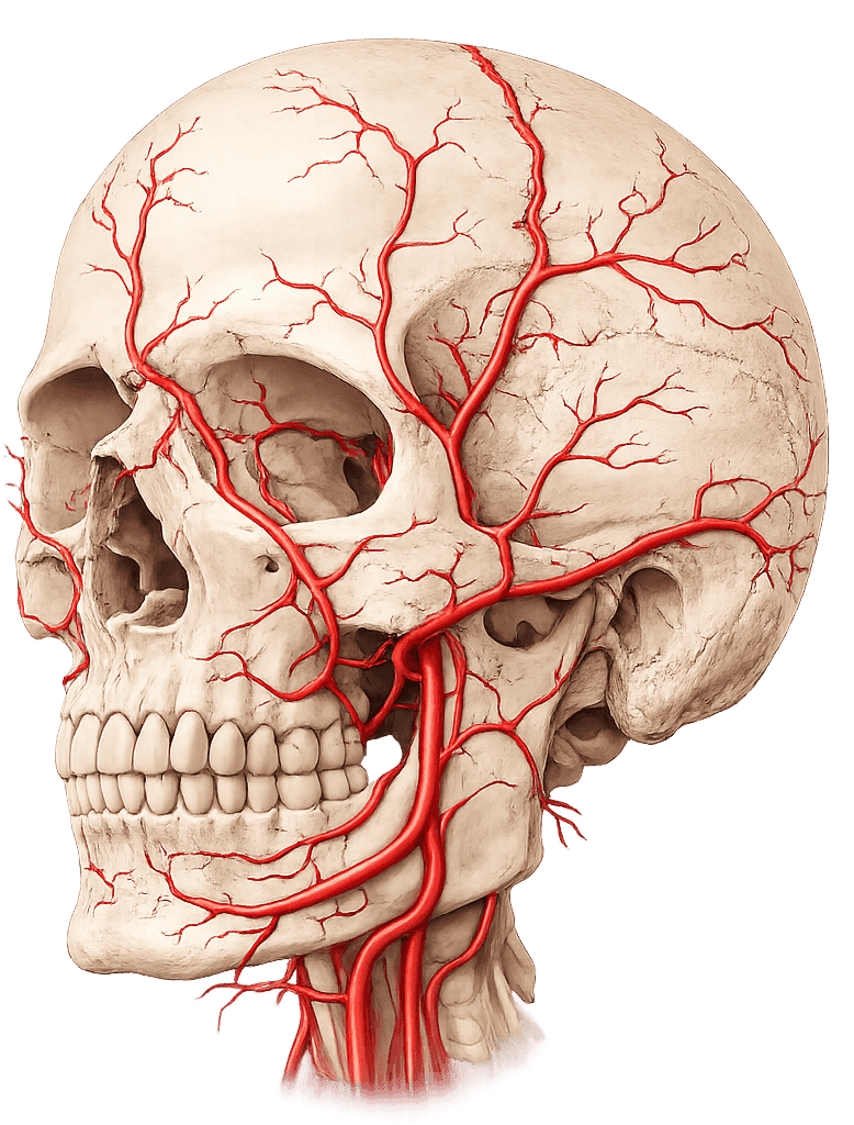
The skull arterial system comprises the external carotid artery, internal carotid artery, vertebrobasilar system, and their principal distribution pathways.
External Carotid Artery
MYO CORE
Core Anatomy
The external carotid artery is the principal extracranial arterial trunk of the head and neck, supplying the craniofacial skeleton, scalp, meninges, upper aerodigestive tract, endocrine structures, and surrounding soft tissues.
OVERVIEW
Functionally – it represents the major conduit through which systemic circulation reaches the superficial and deep extracranial compartments of the head.
Through its extensive branching network and numerous anastomotic channels, the ECA contributes not only to regional tissue perfusion but also to collateral cerebral circulation when carotid or vertebrobasilar blood flow becomes compromised.


ANATOMY
Origin
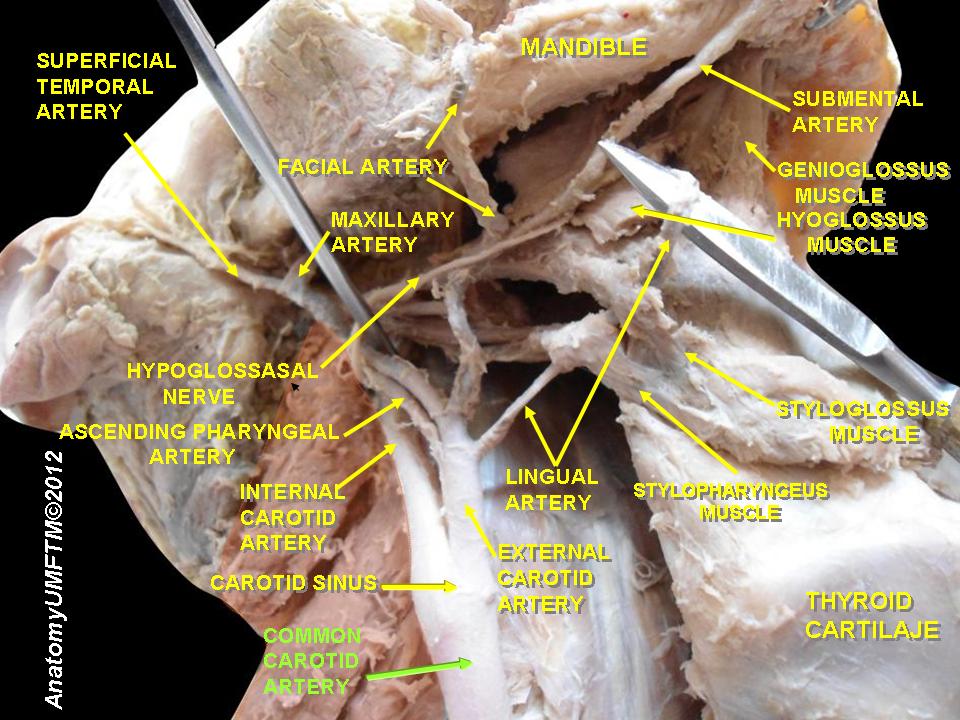
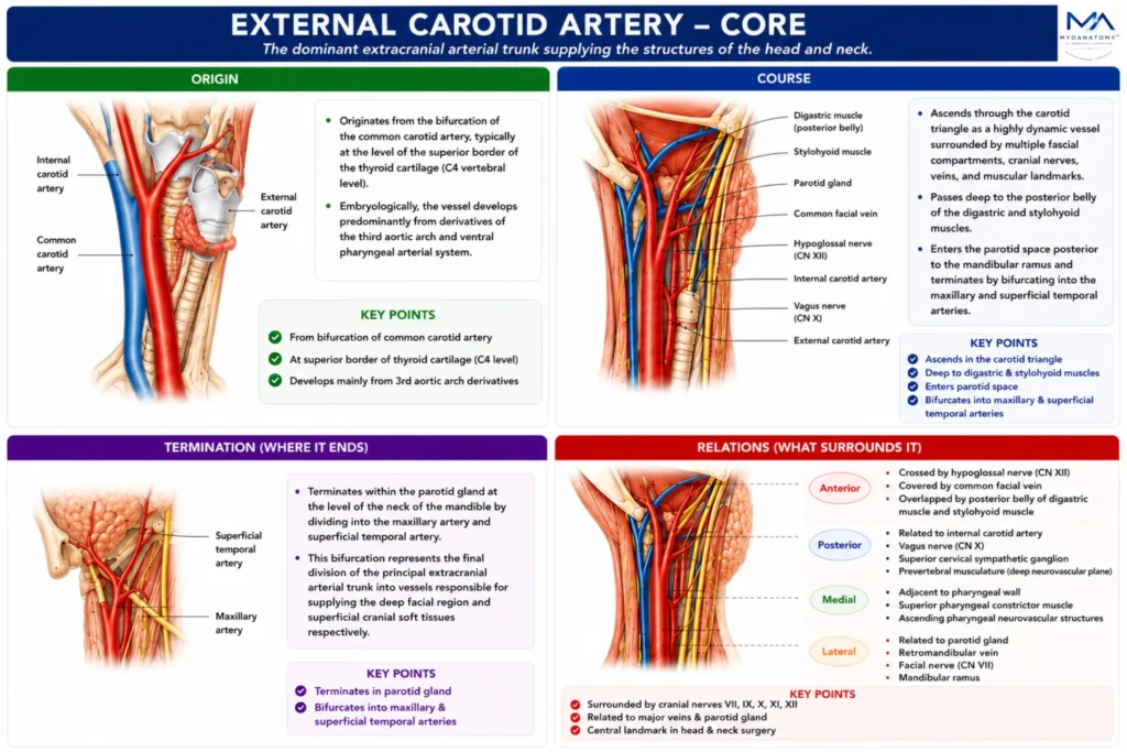
The external carotid artery originates from the bifurcation of the common carotid artery, typically at the level of the superior border of the thyroid cartilage (C4 vertebral level).
Embryologically, the vessel develops predominantly from derivatives of the third aortic arch and ventral pharyngeal arterial system, explaining its intimate association with structures derived from the pharyngeal arches.
At its origin, the ECA occupies an anteromedial position relative to the internal carotid artery; however, progressive rotational changes during cervical ascent result in the artery assuming a more anterior and lateral relationship.
The carotid bifurcation constitutes a major hemodynamic transition point where blood flow is distributed between intracranial and extracranial circulatory territories.
Exam Question
Analyze the anatomical, embryological, and hemodynamic significance of the origin of the external carotid artery at the common carotid bifurcation, including its relationship to the internal carotid artery during cervical ascent.
Course
The external carotid artery ascends through the carotid triangle as a highly dynamic vascular structure surrounded by multiple fascial compartments, cranial nerves, venous channels, and muscular landmarks. Unlike the internal carotid artery, which remains branchless within the neck, the ECA continuously distributes collateral branches during its ascent.
As it courses superiorly, the vessel passes deep to the posterior belly of the digastric and stylohyoid muscles, structures that mark the transition between the carotid and suprahyoid regions.
The artery subsequently enters the parotid space posterior to the mandibular ramus, where it becomes embedded within the substance of the parotid gland. Within this glandular compartment, the ECA forms part of a complex neurovascular arrangement involving the facial nerve, retromandibular vein, and terminal arterial branches.
Exam Question
Describe the complete cervical course of the external carotid artery from its origin to entry into the parotid gland, emphasizing its relationships to the digastric and stylohyoid muscles, carotid space, and surrounding neurovascular structures.
Termination
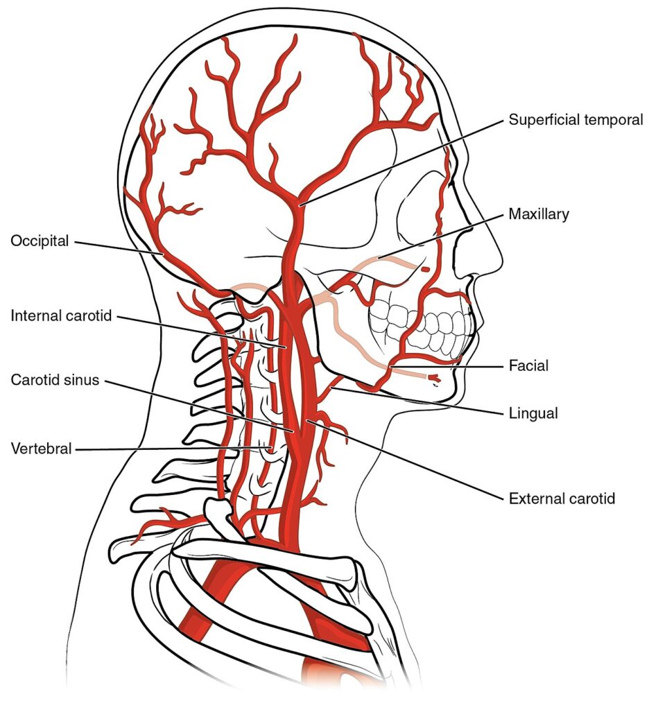
The external carotid artery terminates within the parotid gland at the level of the neck of the mandible by dividing into the maxillary artery and superficial temporal artery.
This bifurcation represents the final division of the principal extracranial arterial trunk into vessels responsible for supplying the deep facial region and superficial cranial soft tissues respectively.
The maxillary artery continues as the dominant arterial supply of the infratemporal fossa, nasal cavity, oral cavity, teeth, meninges, and deep facial structures.
The superficial temporal artery ascends over the zygomatic arch to vascularize the scalp and temporal region. Together these terminal branches form critical components of the craniofacial arterial network and participate extensively in collateral vascular pathways
Exam Question
Discuss the anatomical and functional significance of the terminal bifurcation of the external carotid artery into the maxillary and superficial temporal arteries, and explain how these vessels contribute to craniofacial vascularization.
Relations
Anteriorly – it is crossed by the hypoglossal nerve (CN XII), common facial vein, lingual vein, posterior belly of the digastric muscle, and stylohyoid muscle, which serve as important landmarks within the carotid triangle.
Posteriorly- it is related to the internal carotid artery, vagus nerve (CN X), superior cervical sympathetic ganglion, and prevertebral musculature, forming part of the deep neurovascular plane of the neck.
Medially- the vessel lies adjacent to the pharyngeal wall, superior pharyngeal constrictor muscle, and ascending pharyngeal neurovascular structures, reflecting its close association with the upper aerodigestive tract.
Laterally- it becomes related to the parotid gland, retromandibular vein, facial nerve (CN VII), and mandibular ramus. Within the parotid gland, the artery forms an important component of the gland’s neurovascular architecture.
Because of its intimate relationships with cranial nerves VII, IX, X, XI, and XII, major venous channels, and cervical fascial compartments, the external carotid artery represents a central anatomical landmark in head and neck surgery.

Exam Question
Evaluate the surgical importance of the anatomical relations of the external carotid artery, including its associations with cranial nerves, major veins, cervical fascial compartments, and the parotid gland.
SUMMARY TABLE