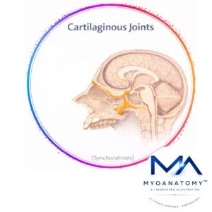
Cartilaginous Joint
MYO CORE
Sphenoethmoidal Synchondrosis
The spheno-ethmoidal synchondrosis is the primary organizer of anterior cranial base development, integrating facial, orbital, and nasal growth through early endochondral expansion.
OVERVIEW
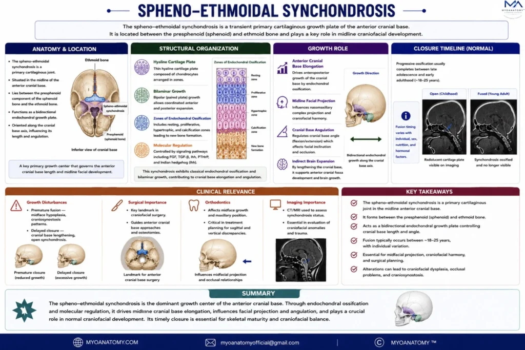
The spheno-ethmoidal synchondrosis is a transient primary cartilaginous growth plate of the anterior cranial base, located between the presphenoid component of the sphenoid and the ethmoid bone.
It represents a critical interface within the chondrocranium where endochondral ossification coordinates anterior cranial base elongation with the morphogenesis of the naso-orbital complex.
Unlike the spheno-occipital synchondrosis, its functional significance is front-loaded in early development, where it establishes the anterior craniofacial framework upon which later growth is superimposed.
Exam Question
Why is the spheno-ethmoidal synchondrosis considered a transient anterior cranial base growth center, and how does its early activity establish the foundational naso-orbital craniofacial framework?
ANATOMY
Anatomical Position
This synchondrosis lies along the midline anterior cranial base, immediately posterior to the cribriform plate and ethmoidal labyrinth, and anterior to the presphenoid body forming part of the sella region.
Superiorly, it contributes to the floor of the anterior cranial fossa, supporting the frontal lobes, while inferiorly it is contiguous with the nasal septal cartilage and superior nasal cavity roof.
Laterally, it is functionally related to the medial orbital walls, placing it at a convergence point between neurocranial and viscerocranial domains
Exam Question
How does its position between the cribriform plate, presphenoid, and ethmoidal labyrinth functionally integrate the anterior cranial fossa with the nasal and orbital complexes?
Structural Organization
Histologically, the spheno-ethmoidal synchondrosis is composed of hyaline cartilage arranged as a bipolar growth plate, though less robust than posterior synchondroses. It demonstrates classical zones of endochondral ossification, including:
resting chondrocytes maintaining matrix integrity
proliferative chondrocyte columns aligned along the cranial base axis
hypertrophic chondrocytes undergoing volumetric expansion
zones of matrix calcification and vascular invasion
Cellular activity is regulated by tightly controlled molecular gradients, including FGF signaling (particularly FGFR mutations in craniosynostosis syndromes), TGF-β pathways, and SHH (sonic hedgehog) signaling, which is especially critical in midline craniofacial patterning.
Functionally, this synchondrosis exhibits predominantly unidirectional growth, oriented anteriorly, contributing to expansion of the anterior cranial base rather than global cranial base elongation.
Exam Question
How does its bipolar hyaline cartilage organization with classical endochondral zones support predominantly unidirectional anterior growth, and how is this distinct from posterior synchondroses?
Growth Role
The spheno-ethmoidal synchondrosis plays a decisive role in early craniofacial spatial organization, acting as an anterior growth regulator that determines:
anteroposterior positioning of the ethmoid complex, influencing nasal septum development
vertical and horizontal alignment of the orbital cavities, particularly the medial walls and orbital roof
projection and angulation of the nasofrontal region, contributing to the craniofacial profile
Through these mechanisms, it indirectly modulates the relationship between the anterior cranial fossa and facial skeleton, ensuring that brain expansion anteriorly is matched by appropriate facial projection and nasal cavity development.
Importantly, growth at this synchondrosis establishes early cranial base length anterior to the sella, which subsequently influences facial growth vectors and midfacial proportions.
Exam Question
How does growth at this synchondrosis regulate anteroposterior positioning of the ethmoid, orbital alignment, and nasofrontal angulation, thereby determining early craniofacial spatial organization?
Closure
The spheno-ethmoidal synchondrosis undergoes early fusion relative to posterior cranial base growth centers, typically ossifying during early childhood ( 7 years old).
This early closure reflects its role in initial craniofacial patterning rather than sustained postnatal growth modulation, after which growth shifts predominantly to sutural and posterior synchondrosal activity.
Exam Question
What developmental mechanisms explain its early ossification in childhood (~7 years), and how does this reflect its role in initial craniofacial patterning rather than sustained growth?
Clinical Relevance
Although less clinically conspicuous than the spheno-occipital synchondrosis, disturbances in this region have profound developmental implications, particularly because they occur during critical windows of craniofacial patterning.
Pathological alterations may lead to:
midline craniofacial dysplasia, due to disrupted SHH-dependent patterning
nasal septal deviations and underdevelopment, affecting airway architecture
orbital asymmetry or hypertelorism/hypotelorism, due to altered ethmoidal and medial orbital positioning
anterior cranial base shortening, influencing facial projection
In syndromic craniosynostoses (e.g., FGFR-related disorders), abnormal signaling at this synchondrosis contributes to complex craniofacial phenotypes involving the orbitonasal region.
From a clinical imaging perspective, this region is relevant in early developmental assessment, particularly in pediatric neuroradiology, where subtle abnormalities may indicate global craniofacial growth disturbances.
SUMMARY TABLE