The auricular muscles are a small group of muscles of facial expression associated with movement of the external ear (auricle). They consist of three extrinsic muscles: auricularis anterior; auricularis superior; auricularis posterior. These muscles connect the auricle to the scalp and temporal region, allowing limited movements of the ear. 

Auricularis Muscle

CORE

Overview

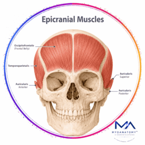
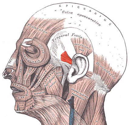
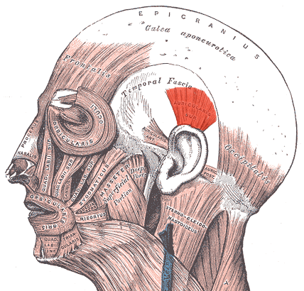
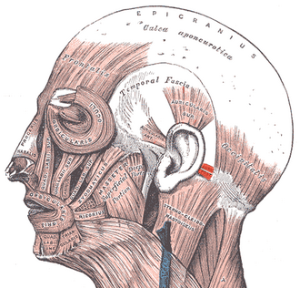
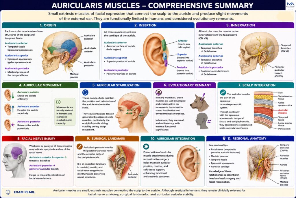
The auricular muscles are small extrinsic muscles that connect the auricle to the scalp and permit slight movements of the external ear. They consist of the auricularis anterior, superior, and posterior muscles and lie within the superficial fascia of the temporal and auricular regions.

In many mammals these muscles are highly developed and play an important role in sound localization through active ear movement. In humans, they are rudimentary and produce only minimal auricular motion, reflecting the reduced functional importance of ear mobility.

Embryologically, they arise from the second pharyngeal arch and are innervated by branches of the facial nerve (CN VII), linking them to the muscles of facial expression. Although functionally limited, they remain anatomically significant because of their close relationships with the facial nerve, scalp fascia, auricle, and surgical anatomy of the temporal and mastoid regions.

ANATOMY

Origin

Each auricular muscle arises from structures of the scalp or temporal fascia. Origin:

Auricularis anterior – temporal fascia and epicranial aponeurosis

Auricularis superior – epicranial aponeurosis (galea aponeurotica)

Auricularis posterior – mastoid process of the temporal bone

Exam Question

Describe the origins of the auricular muscles and explain how their attachments connect the scalp to the external ear.

Insertion

The muscles insert into the cartilage of the auricle.

Auricularis anterior – anterior surface of the auricle (helix region)

Auricularis superior – superior portion of the auricle

Auricularis posterior– posterior surface of the auricle

Exam Question

Explain the insertions of the auricular muscles and how these attachments determine auricular movement.

Innervation

All auricular muscles receive motor innervation from the facial nerve (cranial nerve VII).

Specifically:

Auricularis anterior – temporal branches of the facial nerve

Auricularis superior – temporal branches of the facial nerve

 Auricularis posterior – posterior auricular branch of the facial nerve

The posterior auricular nerve arises from the facial nerve shortly after it exits the stylomastoid foramen.

Exam Question

Identify the facial nerve branches supplying the auricular muscles and discuss the effects of their injury.

FUNCTIONAL ROLE

Auricular Movement

The auricular muscles produce subtle movements of the external ear. The auricularis anterior draws the auricle anteriorly, the auricularis superior elevates it superiorly, and the auricularis posterior retracts it posteriorly. Although these movements are usually minimal in humans, they represent the residual motor capacity of the auricular musculature.

Exam Question

How do the anterior, superior, and posterior auricular muscles contribute to movement of the auricle?

Auricular Stabilization

The auricular muscles help maintain the position and orientation of the auricle relative to the scalp. They counterbalance tension generated by adjacent scalp muscles, particularly the occipitofrontalis, and contribute to the stabilization of the external ear during scalp movement.

Exam Question

Explain how the auricular muscles help maintain the position and orientation of the auricle during scalp movement.

Evolutionary Remnant

In many mammals, the auricular muscles are highly developed and permit independent ear movements that enhance sound localization and environmental awareness. In humans, auricular mobility is greatly reduced, with auditory localization relying primarily on binaural hearing and head movements. 

Consequently, these muscles are regarded as vestigial remnants of a once highly functional auditory-orienting system

Exam Question

Why are the auricular muscles considered vestigial in humans, and what is their functional significance in other mammals?

Scalp Integration

The auricular muscles function as components of the epicranial musculoaponeurotic system. Through their continuity with the epicranial aponeurosis, temporal fascia, and occipitofrontalis muscle, they participate in coordinated scalp–auricular mechanics. 

This integration assists in maintaining auricular alignment, transmitting fascial tension, and preserving the structural orientation of the external ear.

Exam Question

Discuss the relationship between the auricular muscles, galea aponeurotica, temporal fascia, and occipitofrontalis.

CLINICAL RELEVANCE

Facial Nerve Injury

Because the auricular muscles receive motor innervation from branches of the facial nerve (CN VII), their function may be impaired in facial nerve palsy.

Although loss of auricular movement is rarely clinically significant, weakness of these muscles may indicate injury to the temporal or posterior auricular branches of the facial nerve and can assist in the anatomical localization of facial nerve lesions.

Exam Question

How can weakness of the auricular muscles assist in the localization of facial nerve lesions?

Surgical Landmark

The auricular muscles, particularly the auricularis posterior, serve as important landmarks during mastoid, parotid, and facial nerve surgery.

Their close relationship to the posterior auricular nerve aids surgeons in identifying and preserving critical neural structures. The posterior auricular nerve is especially important because it also supplies the occipital belly of the occipitofrontalis muscle.

Exam Question

Why is the auricularis posterior an important landmark during mastoid, parotid, and facial nerve surgery?

Auricular Integration

Detailed knowledge of auricular muscle anatomy is important during reconstructive procedures involving the external ear. 

Preservation of the surrounding muscular and fascial attachments helps maintain normal auricular position, contour, and soft-tissue support, contributing to improved functional and aesthetic outcomes.

Exam Question

Discuss the importance of preserving auricular muscular attachments during external ear reconstruction.

Regional Anatomy

Despite their limited motor role, the auricular muscles remain clinically relevant because of their intimate relationships with the facial nerve, scalp fascia, mastoid region, and auricular cartilage. Consequently, they serve as valuable anatomical reference structures in head and neck surgery, facial reanimation procedures, and auricular reconstruction.

Exam Question

Describe the key anatomical relationships of the auricular muscles and explain their clinical significance in head and neck surgery.

SUMMARY TABLE

Scroll to Top