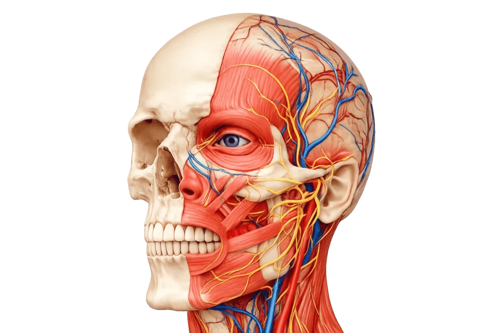
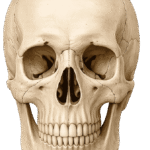
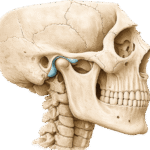
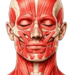
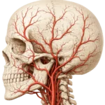
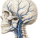
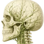
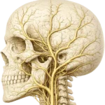
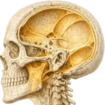
Skull Anatomy Cranial Bones, Facial Structures and key Neurovascular Pathways Explore the intricate architecture of the skull, including its bones, joints, muscles and the vital neurovascular structures that support and protect. Skull Bones Neurocranium Forms the protective cranial vault enclosing the brain and providing attachment for the meninges and cranial muscles. Viscerocranium Comprises the facial skeleton, supporting the orbits, nasal cavity, oral cavity, and upper airway. Orbital Cavity A pyramidal bony cavity housing the eyeball, extraocular muscles, cranial nerves, and orbital vessels. Nasal Cavity A complex osteocartilaginous chamber responsible for air conditioning, olfaction, and respiratory filtration. Explore Skull Joints Synovial Joints The temporomandibular joint, the only synovial articulation of the skull, enabling mastication and speech. Cartilaginous Joints Primary synchondroses of the cranial base that contribute to skull development and endochondral growth. Cranial Sutures Fibrous joints that unite the cranial bones, permitting skull growth before eventually becoming largely immobile. Explore Skull Muscles Facial Muscles Muscles inserting into the skin to produce facial movements and non-verbal communication. Masticatory Muscles Powerful muscles responsible for mandibular elevation, depression, protrusion, and lateral excursion during chewing. Extraocular Muscles Six muscles precisely controlling ocular alignment, gaze, and coordinated eye movements. Suprahyoid Muscles Muscles connecting the mandible and hyoid, assisting swallowing, speech, and mandibular depression. Explore Skull Arteries External Carotid Supplies the scalp, face, jaws, meninges, and most superficial structures of the head. Internal Carotid Provides the principal arterial supply to the anterior cerebral circulation and orbital contents. Vertebro Basilar Supplies the posterior brain, brainstem, cerebellum, and contributes to the cerebral arterial circle. Explore Skull Veins Superficial Veins Drain the scalp and face into the external and internal jugular venous systems. Deep Veins Drain deep facial structures, communicating with intracranial venous channels through emissary pathways. Dural Sinuses Endothelial venous channels within the dura mater collecting blood and cerebrospinal fluid from the brain. Explore Skull Lymphatics Superficial Nodes Receive lymph from the scalp, face, and superficial tissues of the head. Facial Nodes Drain the facial soft tissues before emptying into the deep cervical lymphatic chain. Deep Cervical Nodes Principal lymphatic drainage stations for the head and neck, receiving lymph from superficial and deep structures. Explore Cranial Nerves CN I-XII Classification The twelve cranial nerves are classified as sensory, motor, or mixed according to their functional fiber composition. Fiber Modalities Functional fiber types transmit somatic, visceral, special sensory, and autonomic information between the brain and target tissues. Brainstem Nuclei Specialized nuclei within the brainstem serve as the origin or termination of cranial nerve fibers. Course & Distribution Each cranial nerve exits the cranial cavity through specific foramina to reach its peripheral distribution. Explore Cranial Spaces Cranial Foramina Openings within the skull that transmit cranial nerves, arteries, veins, and other neurovascular structures. Cranial Fossae Three intracranial compartments supporting the cerebral hemispheres, cerebellum, and brainstem. Pterygopalatine Fossae A neurovascular crossroads transmitting branches of the maxillary nerve and terminal maxillary artery. Infratemporal Fossae A deep anatomical space containing the muscles of mastication and major neurovascular structures. Explore Deep Spaces Masticator Space A deep fascial compartment containing the muscles of mastication, mandibular ramus, and mandibular nerve branches. Parapharyngeal Space A central neck space transmitting major vessels, cranial nerves, and lymphatic structures. Retropharyngeal Space A potential fascial space posterior to the pharynx that permits neck movement but may facilitate deep infection spread Explore Key Point All muscles of the Facial expression are innervated by the facial nerve (CN VII), except for the Platysma, which receives innervation from the cervical branch of the facial nerve (CN VII).