Bone Anatomy
Bone anatomy explores the structure, development, classification, and lifelong adaptation of bones that underpin skeletal function and biomechanical integrity.

Bone Classification
MYO CORE
Oveview
Bone classification is a fundamental concept in osteology that organizes bones according to their morphology, internal structure, and functional role within the musculoskeletal system.This framework provides a basis for understanding how skeletal elements are adapted to meet specific mechanical demands, including load transmission, protection, and movement facilitation.
OVERVIEW
The structural form of a bone is closely related to its function, reflecting the principle that architecture follows biomechanics, where variations in shape and composition correspond to differences in mechanical stress distribution and functional requirements.
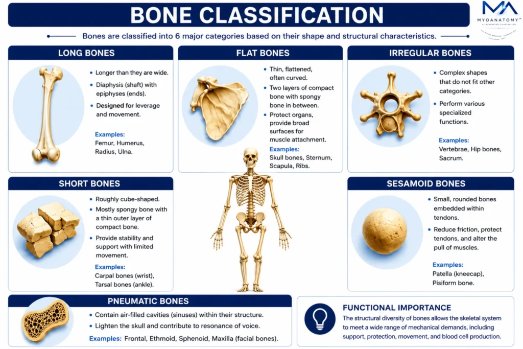
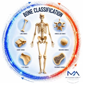
Bones are classified into 6 major categories based on their shape and structural characteristics: long bones, short bones, flat bones, irregular bones, sesamoid bones, and pneumatic bones. Each category exhibits distinct structural adaptations that optimize performance for specific roles, such as leverage, stability, protection, or force modulation.
These classifications are essential for interpreting skeletal organization, joint mechanics, and muscular attachment patterns, as well as for understanding how structural variations influence biomechanical behavior across the musculoskeletal system.
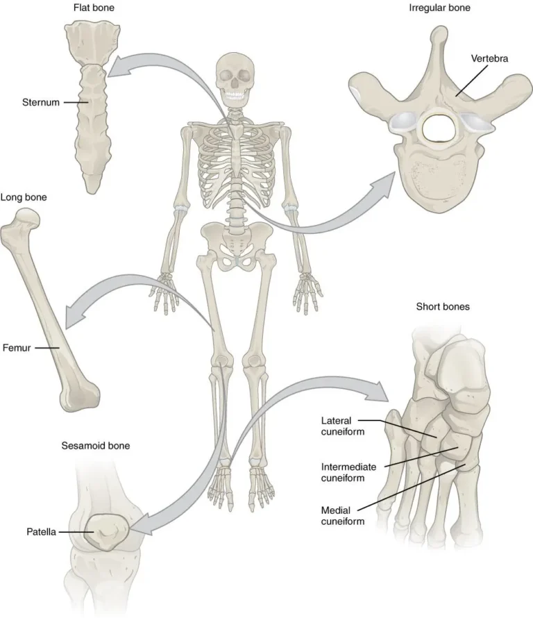
”Bone classification” by OpenStax College, via Wikimedia Commons. Licensed under CC BY SA 3.0
ANATOMY
Long Bones
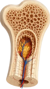
Long bones are defined by a length exceeding their width, characterized by a cylindrical shaft (diaphysis) and expanded ends (epiphyses). Structurally, the diaphysis consists predominantly of compact (cortical) bone surrounding a medullary cavity, while the epiphyses are composed mainly of trabecular (spongy) bone, optimized for load dispersion.
Functionally, long bones act as mechanical levers, facilitating movement by transmitting muscular forces across joints and providing extensive sites for muscle attachment. Their elongated architecture enables efficient force amplification and directional control of motion.
Examples: humerus, radius, ulna, femur, tibia, fibula, metacarpals, metatarsals, and phalanges.
From a biomechanical perspective, long bones are essential for locomotion and limb dynamics, forming the structural framework through which muscles generate controlled movement, particularly within synovial joint systems.
Exam Question
In a patient with impaired limb movement, which structural and functional characteristics of long bones explain their role in force transmission, leverage, and controlled joint motion?
Short Bones
Short bones are approximately cuboidal in shape, with similar dimensions in length, width, and thickness. They consist of a core of trabecular bone enclosed by a thin shell of compact bone, providing structural strength with minimal weight.
These bones are located in regions requiring stability with controlled mobility, where their architecture allows efficient distribution of compressive forces while permitting limited gliding movements between adjacent bones.
Examples: carpal bones of the wrist and the tarsal bones of the ankle.
Functionally, short bones play a key role in shock absorption, load distribution, and fine-tuned motion control, particularly within complex joint systems such as the wrist and ankle
Exam Question
In the context of wrist biomechanics, which structural features of short bones account for their ability to provide stability, shock absorption, and controlled gliding movements under compressive loads?
Flat Bones
Flat bones are thin, flattened structures composed of two layers of compact bone enclosing an inner layer of trabecular (spongy) bone, known as the diploë, which enhances structural strength while maintaining reduced weight. This layered architecture provides resistance to bending forces and distributes mechanical stress across a broad surface area.
Functionally, flat bones serve two principal roles: protection of vital organs and provision of extensive surfaces for muscular attachment. Their broad geometry allows efficient force distribution and supports large muscle groups involved in stabilization and coordinated movement of major skeletal regions
Examples: cranial vault, scapula , sternum and ribs.
From a biomechanical perspective, flat bones contribute to structural protection and force dispersion, particularly within regions such as the cranial vault, thoracic cage, and shoulder girdle, where protection and stability are critical.
Exam Question
How does the layered structural organization of flat bones (compact–trabecular–compact) explain their role in mechanical stress distribution, resistance to bending forces, and protection of vital organs?
Irregular Bones
Irregular bones are characterized by complex, non-uniform shapes that do not conform to standard geometric categories. Their structural configuration reflects highly specialized adaptations for mechanical support and protection, allowing them to accommodate diverse functional demands.
Within the musculoskeletal system, irregular bones serve as critical sites for muscle attachment, provide structural stability, and contribute to the protection of essential neural elements, particularly the spinal cord. Their architecture enables multidirectional load handling and supports complex biomechanical interactions.
Examples: vertebrae, sacrum, coccyx, many bones of the skull.
Functionally, irregular bones are integral to maintaining postural stability, load transmission, and protection of neural structures, especially within the vertebral column and craniofacial skeleton.
Exam Question
How does the complex, non-uniform architecture of irregular bones support their roles in multidirectional load handling, muscle attachment, and protection of neural structures within the musculoskeletal system?
Sesamoid Bones
Sesamoid bones are small, rounded bones that develop within tendons, typically at sites where tendons pass over joints and are subjected to significant mechanical stress. Their presence modifies the direction of tendon pull, reducing friction and protecting tendons from excessive wear against adjacent structures.
Example: patella represents the largest sesamoid bone, embedded within the quadriceps tendon, playing a critical role in knee extension mechanics.
Functionally, sesamoid bones enhance mechanical efficiency by increasing the moment arm of muscles, thereby improving force transmission and optimizing the force-generating capacity of the musculoskeletal system.
Exam Question
How does the development of sesamoid bones within tendons modify mechanical leverage, tendon stress distribution, and force transmission across joints during movement?
Pneumatic Bones
Pneumatic bones are characterized by the presence of air-filled cavities (sinuses) within their structure, which are lined by mucous membrane and communicate with the respiratory tract. These cavities reduce bone mass while preserving structural integrity.
Functionally, pneumatic bones contribute to weight reduction of the skull without compromising strength, thereby optimizing load efficiency for head support. Additionally, they play a role in resonance of voice and participate in the overall biomechanical balance of craniofacial structures.
Example: several bones of the skull, such as frontal, sphenoid, ethmoid and maxilla.
From a structural perspective, this adaptation allows maintenance of mechanical strength with minimal mass, which is critical for efficient musculoskeletal support of the head.
Exam Question
How does the presence of air-filled cavities within pneumatic bones enable reduction of skeletal mass while preserving structural integrity and contributing to craniofacial function?
SUMMARY TABLE