Synovial Joint
MYO CORE
Morphology
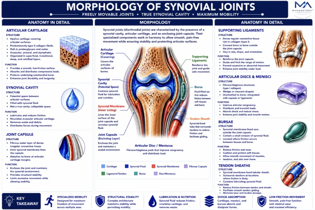
Synovial joints are highly specialized articulations whose morphology is defined by an integrated structural complex consisting of articular cartilage, a synovial cavity, a fibrous capsule, supporting ligaments, fibrocartilaginous adaptations (discs and menisci), and accessory synovial structures such as bursae and tendon sheaths. Together, these components optimize mobility, stability, lubrication, load distribution, and mechanical efficiency while preserving joint integrity under dynamic physiological loading.
OVERVIEW
Synovial joints represent the most specialized and mobile class of joints within the human musculoskeletal system. Their morphology is characterized by the presence of a synovial cavity, articular cartilage, a fibrous capsule, supporting ligaments, and accessory structures that function as an integrated biomechanical unit.
Together, these components minimize friction, facilitate smooth movement, distribute mechanical loads, absorb shock, and maintain joint stability under dynamic physiological conditions. The structural organization of synovial joints allows an exceptional range of motion while preserving the integrity of articular surfaces during lifelong functional activity.
Consequently, synovial joints form the anatomical foundation for locomotion, manipulation, posture, and complex human movement, representing the highest degree of mobility within the skeletal system.
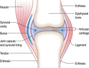
Synovial Joint Example (Joint.png)” – Madhero88, via Wikimedia Commons. Licensed under CC BY SA 3.0
How does the integrated structural organization of synovial joints enable a balance between mobility, stability, load distribution, and long-term preservation of articular integrity?
Exam Question
ANATOMY
Articular Cartilage
Articular cartilage in synovial joints is a specialized form of hyaline cartilage composed predominantly of type II collagen fibrils embedded within a highly hydrated proteoglycan-rich extracellular matrix. Chondrocytes are sparsely distributed within lacunae and maintain matrix homeostasis through tightly regulated synthesis and degradation.
This tissue is avascular, aneural, and alymphatic, relying entirely on diffusion from synovial fluid for nutrient supply and waste removal. It exhibits a zonal organization (superficial, transitional, deep, and calcified layers), each adapted to specific mechanical functions, including shear resistance at the surface and compressive load absorption in deeper regions.
The extracellular matrix provides viscoelastic properties, allowing deformation under load and recovery upon unloading, thereby distributing forces efficiently and protecting the underlying subchondral bone while maintaining a low-friction articulating surface.
Exam Question
Explain how the zonal architecture and extracellular matrix composition of articular cartilage enable it to withstand both compressive and shear forces while remaining avascular.
Synovial Cavity
The synovial cavity is a fluid-filled space separating the articular surfaces, essential for frictionless joint movement. It contains synovial fluid, a plasma-derived ultrafiltrate enriched with hyaluronic acid and lubricin, which provides lubrication, reduces wear, and facilitates nutrient diffusion to avascular articular cartilage.
Synovial fluid exhibits non-Newtonian, shear-thinning behavior, allowing it to adapt to varying mechanical demands – becoming less viscous during rapid movement to reduce friction, and more viscous at rest to enhance joint stability.
The cavity is enclosed by a fibrous capsule lined internally by the synovial membrane, which regulates fluid composition and maintains joint homeostasis. Through continuous circulation and pressure changes during movement, synovial fluid supports metabolic exchange and contributes to shock absorption and load distribution.
Exam Question
Analyze how the biochemical composition and rheological properties of synovial fluid contribute to both lubrication and nutrition of articular cartilage in synovial joints
Articular Cartilage
Articular cartilage in synovial joints is a specialized form of hyaline cartilage composed predominantly of type II collagen fibrils embedded within a highly hydrated proteoglycan-rich extracellular matrix. Chondrocytes are sparsely distributed within lacunae and maintain matrix homeostasis through tightly regulated synthesis and degradation.
This tissue is avascular, aneural, and alymphatic, relying entirely on diffusion from synovial fluid for nutrient supply and waste removal. It exhibits a zonal organization (superficial, transitional, deep, and calcified layers), each adapted to specific mechanical functions, including shear resistance at the surface and compressive load absorption in deeper regions.
The extracellular matrix provides viscoelastic properties, allowing deformation under load and recovery upon unloading, thereby distributing forces efficiently and protecting the underlying subchondral bone while maintaining a low-friction articulating surface.
Exam Question
Explain how the zonal architecture and extracellular matrix composition of articular cartilage enable it to withstand both compressive and shear forces while remaining avascular.
Synovial Cavity
The synovial cavity is a fluid-filled space separating the articular surfaces, essential for frictionless joint movement. It contains synovial fluid, a plasma-derived ultrafiltrate enriched with hyaluronic acid and lubricin, which provides lubrication, reduces wear, and facilitates nutrient diffusion to avascular articular cartilage.
Synovial fluid exhibits non-Newtonian, shear-thinning behavior, allowing it to adapt to varying mechanical demands – becoming less viscous during rapid movement to reduce friction, and more viscous at rest to enhance joint stability.
The cavity is enclosed by a fibrous capsule lined internally by the synovial membrane, which regulates fluid composition and maintains joint homeostasis. Through continuous circulation and pressure changes during movement, synovial fluid supports metabolic exchange and contributes to shock absorption and load distribution.
Exam Question
Analyze how the biochemical composition and rheological properties of synovial fluid contribute to both lubrication and nutrition of articular cartilage in synovial joints
Joint Capsule
Each synovial joint is enclosed by a joint capsule forming a continuous sleeve between adjacent bones and blending with the periosteum. It consists of two structurally and functionally distinct layers.
The outer fibrous layer is composed of dense irregular connective tissue rich in type I collagen, arranged to resist multidirectional mechanical stresses. It provides tensile strength, structural containment, and limits excessive joint movement.
The inner synovial membrane is a specialized connective tissue lacking a true epithelium and containing synoviocytes. Type A synoviocytes perform phagocytic functions, while Type B synoviocytes synthesize synovial fluid components, including hyaluronic acid and lubricin, maintaining the biochemical environment of the joint.
This dual-layered organization ensures both mechanical stability and metabolic regulation of the intra-articular space, enabling efficient movement while preserving joint integrity.
Exam Question
Explain how the structural differences between the fibrous capsule and synovial membrane contribute to both mechanical stability and metabolic homeostasis in synovial joints.
Supporting Ligaments
Ligaments are dense connective tissue structures that reinforce synovial joints and integrate with surrounding musculoskeletal components to guide and constrain movement.
They are classified based on their anatomical relationship to the joint capsule: capsular ligaments (thickenings of the fibrous capsule), extracapsular ligaments (external to the capsule), and intracapsular ligaments (located within the capsule but excluded from the synovial cavity).
Ligaments are primarily composed of type I collagen fibers arranged in parallel bundles aligned with dominant mechanical stress vectors. This organization allows controlled elongation under tension while resisting excessive displacement, contributing to joint stability and directional control of movement.
Functionally, ligaments act as passive stabilizers, limiting abnormal motion, maintaining joint congruency, and providing proprioceptive feedback through embedded mechanoreceptors.
Exam Question
Analyze how the collagen fiber orientation and anatomical positioning of ligaments determine their role in guiding joint motion and preventing instability.
Articular Disk & Menisci
Certain synovial joints contain fibrocartilaginous intra-articular structures – articular discs and menisci – composed predominantly of type I collagen arranged to resist multidirectional mechanical stresses. These structures may partially or completely divide the synovial cavity, creating functionally distinct compartments within a single joint.
Their morphology is joint-specific: crescent-shaped menisci in the knee enhance congruency between incongruent surfaces, while complete discs (e.g., temporomandibular joint) allow complex combined movements. The organized collagen architecture enables resistance to compressive, tensile, and shear forces.
Functionally, discs and menisci optimize load distribution, increase joint stability, guide movement, and reduce focal stress by improving articular congruency and dissipating mechanical energy across the joint surface.
Exam Question
Explain how the fibrocartilaginous composition and structural organization of articular discs and menisci contribute to load distribution and joint stability in synovial joints.
Bursae & Tendon Sheets
Bursae are discrete synovial fluid-filled sacs located at sites of friction between soft tissues and bone or adjacent structures. They are lined by a synovial membrane-like layer and contain a thin film of lubricating fluid that reduces mechanical resistance during movement.
Tendon sheaths are specialized elongated bursae that envelop tendons, particularly where they traverse confined spaces or pass over osseous prominences. They facilitate smooth gliding of tendons while maintaining alignment and reducing wear.
Together, these structures form an integrated system of friction-reducing interfaces within the musculoskeletal system, minimizing energy loss, preventing tissue damage, and enabling efficient, repetitive movement.
Exam Question
Analyze how bursae and tendon sheaths contribute to reducing friction and preserving mechanical efficiency in regions of high repetitive motion.
SUMMARY TABLE