
Muscle Classification
Muscle classification in general myology provides a systematic framework for relating skeletal muscle architecture to biomechanical function. Variations in morphology, fascicular arrangement, and attachment patterns reflect specialization for force generation, excursion, precision, or stabilization. Classification based on structural and functional parameters reveals how muscle design determines mechanical performance and enables coordinated movement within integrated kinetic chains of the musculoskeletal system
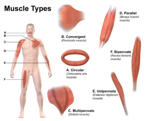
”Muscle Types” – BruceBlaus( Own work), via Wikimedia Commons. Licensed under CC BY-SA 4.0
Definition
A fundamental approach in general myology classifies skeletal muscles according to their external morphology, as muscle shape directly reflects underlying architectural organization and biomechanical function. The geometric configuration of a muscle determines the arrangement of its fibers, tendon orientation, and physiological cross-sectional area, thereby influencing force generation, excursion, and range of motion.
Consequently, muscle morphology represents not merely a descriptive feature but a primary determinant of mechanical performance. Classification based on shape provides critical insight into functional specialization, particularly in the relationship between force production and shortening capacity. The principal morphological categories include parallel, fusiform, convergent, pennate, and circular muscles, each adapted to specific mechanical demands within the musculoskeletal system.
Parallel Muscles
Parallel muscles are characterized by muscle fibers that run parallel to the longitudinal axis of the muscle and to the line of pull between origin and insertion. Because the fibers extend along the length of the muscle, these muscles are capable of considerable shortening during contraction, allowing them to produce large ranges of motion.
Parallel muscles typically exhibit long muscle fibers and relatively smaller physiological cross-sectional areas, meaning they are optimized for movement amplitude rather than maximal force production.
Examples include:
sartorius – the longest muscle in the human body, involved in hip and knee movements
rectus abdominis – responsible for flexion of the vertebral column
biceps brachii – involved in elbow flexion and forearm supination.
Because the fibers run in the same direction as the line of pull, contraction of parallel muscles results in efficient shortening of the entire muscle length, producing substantial joint excursion.
Fusiform Muscles
Fusiform muscles represent a specialized subtype of parallel muscle characterized by a spindle-shaped morphology, with a thickened central muscle belly and tapered ends that attach to tendons.
This architecture allows the muscle to contain relatively long fibers arranged parallel to the tendon, enabling both effective contraction and a moderate capacity for force generation.
Examples include:
biceps brachii
brachioradialis
The fusiform structure allows these muscles to generate smooth and coordinated contractions, making them particularly well suited for movements requiring both strength and flexibility of motion, such as elbow flexion.
Convergent Muscles
Convergent muscles possess a broad origin from which muscle fibers converge toward a single tendon of insertion. This triangular or fan-shaped arrangement allows fibers from different regions of the muscle to pull in slightly different directions.
A classic example is the pectoralis major muscle, which originates broadly from the clavicle, sternum, and ribs before converging into a single tendon inserting on the humerus.
This architectural arrangement provides several functional advantages:
allows different regions of the muscle to be activated independently
enables complex and multidirectional movements
facilitates generation of powerful contractions when all fibers act simultaneously.
Consequently, convergent muscles are capable of producing both versatility of movement and substantial force output, depending on which portion of the muscle is activated.
Pennate Muscles
Pennate muscles are characterized by muscle fibers that attach obliquely to a central tendon, resembling the arrangement of barbs along the shaft of a feather. This configuration increases the number of fibers that can be packed into a given muscle volume.
As a result, pennate muscles have a large physiological cross-sectional area, which allows them to generate greater contractile force compared with parallel muscles of similar size.
Pennate muscles are classified into three structural subtypes.
Unipennate Muscles
In unipennate muscles, fibers attach to one side of a tendon.
example: extensor digitorum longus
This arrangement provides increased force production while maintaining relatively simple fiber orientation.
Bipennate Muscles
In bipennate muscles, fibers attach to both sides of a central tendon, forming a symmetrical feather-like structure.
example: rectus femoris
Bipennate muscles contain a greater number of fibers within the same muscle volume, enabling them to produce stronger contractions.
Multipennate Muscles
Multipennate muscles consist of multiple bipennate units arranged around several tendinous structures.
example: deltoid muscle
This complex architecture allows extremely high force production, although the range of muscle shortening is typically reduced compared with parallel muscles.
Thus, pennate muscles are specialized for powerful contractions rather than large ranges of motion.
Circular Muscles
Circular muscles, also known as sphincter muscles, consist of concentric rings of muscle fibers that surround an anatomical opening or body passage.
When these muscles contract, the diameter of the opening decreases, allowing them to regulate the passage of substances through the structure.
Examples include:
orbicularis oris, which controls movements of the mouth
orbicularis oculi, which closes the eyelids.
Circular muscles therefore play an important role in controlling facial expression, regulating openings of body passages, and protecting sensory organs.
Functional Morphology
The shape and architectural arrangement of skeletal muscles strongly influence their biomechanical capabilities.
Parallel and fusiform muscles are adapted for large ranges of motion and rapid contraction.
Pennate muscles are specialized for generating powerful contractions.
Convergent muscles provide versatile movement through selective fiber activation.
Circular muscles regulate openings and maintain functional control of body orifices.
Consequently, classification based on muscle shape illustrates the close relationship between muscle architecture and biomechanical function, helping explain how different muscles contribute to the complex patterns of movement within the musculoskeletal system.
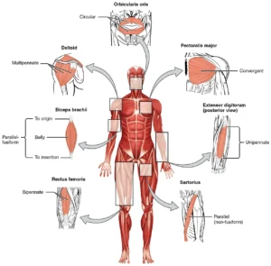
“Skeletal Muscle Types”-OpenStax College, CNX/ via Wikimedia Commons. Licensed under CC BY-SA 3.0
Definition
Another important method of skeletal muscle classification is based on the number of heads or distinct points of origin from which a muscle arises. In this context, the term head refers to an individual portion of a muscle that originates from a separate anatomical structure but ultimately merges with other portions of the muscle to form a common muscle belly or tendon.
Muscles with multiple heads often arise from different bones or anatomical landmarks, allowing them to span multiple joints or contribute to complex patterns of movement. The presence of multiple heads increases the functional versatility and mechanical efficiency of a muscle, as each head may exert slightly different lines of pull depending on its anatomical origin.
This classification system also reflects the structural complexity of muscles and their capacity to participate in multiple actions within the musculoskeletal system.
Several commonly recognized categories exist based on the number of muscle heads.
Biceps Muscles
Biceps Muscles
The term biceps refers to a muscle that possesses two heads of origin. These heads typically arise from separate anatomical structures but converge into a single muscle belly and tendon of insertion.
A classic example is the biceps brachii, which has two heads:
Long head – originates from the supraglenoid tubercle of the scapula
Short head – originates from the coracoid process of the scapula
These two heads merge to form a single muscle belly that inserts on the radial tuberosity and the bicipital aponeurosis.
Functionally, the dual origins allow the muscle to contribute to several actions, including:
elbow flexion
forearm supination
stabilization of the shoulder joint.
The presence of two heads therefore enhances the mechanical versatility and functional efficiency of the muscle.
Triceps Muscles
Triceps muscles possess three heads of origin, which usually arise from different anatomical locations before merging into a common tendon.
The most prominent example is the triceps brachii, the principal extensor muscle of the elbow.
It consists of three heads:
Long head – originates from the infraglenoid tubercle of the scapula
Lateral head – originates from the posterior surface of the humerus above the radial groove
Medial head – originates from the posterior surface of the humerus below the radial groove.
All three heads converge into a common tendon that inserts on the olecranon process of the ulna.
This multi-headed architecture allows the triceps to generate powerful extension of the forearm at the elbow joint, while the long head also contributes to stabilization and extension of the shoulder joint.
Quadriceps Muscles
Quadriceps muscles possess four heads of origin, which combine to form a powerful muscle group responsible for producing large forces during movement.
The primary example is the quadriceps femoris, located in the anterior compartment of the thigh. It consists of four distinct heads:
Rectus femoris
Vastus lateralis
Vastus medialis
Vastus intermedius
These four muscles converge into the quadriceps tendon, which attaches to the patella and continues as the patellar ligament to the tibial tuberosity.
The quadriceps femoris functions primarily to:
extend the knee joint
stabilize the knee during standing and locomotion
assist in hip flexion (rectus femoris).
Because of its four-headed structure, the quadriceps can generate substantial contractile force, making it essential for activities such as walking, running, jumping, and rising from a seated position.
Functional Significance
Classification based on the number of muscle heads highlights how structural complexity enhances muscular function. Muscles with multiple heads often possess:
multiple lines of pull
broader attachment areas
greater capacity for force generation
the ability to participate in movements at more than one joint.
These features allow multi-headed muscles to play key roles in coordinated movement, joint stabilization, and transmission of mechanical forces across the musculoskeletal system.
Consequently, understanding the number and arrangement of muscle heads provides important insight into the anatomical organization, biomechanical performance, and functional versatility of skeletal muscles.
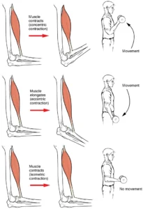
“Types of Muscle Contraction” – OpenStax, Anatomy& Physiology, CNX/Wikimedia Commons. Licensed – under CC BY 4.0
Definition
Skeletal muscles may also be classified according to the type of movement they produce at a joint or the functional role they perform during movement. This functional classification reflects the mechanical actions generated when muscles contract and act upon bones across synovial joints.
Because skeletal muscles rarely function in isolation, functional classification is closely related to the biomechanics of joint movement and the coordination of muscular activity within the musculoskeletal system. Individual muscles typically act in groups to produce complex movements, with different muscles contributing to initiation, stabilization, control, or opposition of motion.
Functional categories of muscles are therefore defined according to the direction of movement they produce relative to anatomical planes and axes of motion.
Flexors Muscles
Flexor muscles produce flexion, a movement that decreases the angle between adjacent skeletal segments. Flexion typically occurs in the sagittal plane around a mediolateral axis.
Examples include:
biceps brachii – flexion of the elbow joint
iliopsoas – flexion of the hip joint
hamstrings – flexion of the knee joint.
Flexor muscles are essential for many daily activities such as grasping, lifting, and forward movement of the limbs.
Extensors Muscles
Extensor muscles produce extension, a movement that increases the angle between articulating bones and often returns a limb toward the anatomical position.
Examples include:
triceps brachii – extension of the elbow joint
quadriceps femoris – extension of the knee joint
gluteus maximus – extension of the hip joint.
Extensors play a critical role in maintaining posture and supporting body weight, particularly in the lower limbs and vertebral column
Abductors Muscles
Abductor muscles move a limb away from the midline of the body, typically within the frontal plane around an anteroposterior axis.
Examples include:
deltoid (middle fibers) – abduction of the shoulder joint
gluteus medius and gluteus minimus – abduction of the hip joint.
Abduction movements are important for positioning the limbs during locomotion and maintaining balance during single-leg stance.
Adductors Muscles
Adductor muscles move a limb toward the midline of the body, opposing the action of abductors.
Examples include:
adductor longus and adductor magnus – adduction of the hip joint
pectoralis major – adduction of the arm at the shoulder.
Adductor muscles help stabilize the limbs during movement and contribute to control of limb positioning during walking, running, and changes in direction.
Rotators Muscles
Rotator muscles produce rotation of a bone around its longitudinal axis, typically occurring in the transverse plane around a vertical axis.
Two primary forms of rotation exist:
medial (internal) rotation – rotation toward the midline
lateral (external) rotation – rotation away from the midline.
Examples include:
subscapularis – medial rotation of the humerus
infraspinatus and teres minor – lateral rotation of the shoulder
gluteus medius (posterior fibers) – lateral rotation of the hip.
Rotational movements are particularly important in ball-and-socket joints such as the shoulder and hip, where large ranges of motion are required.
Pronators Muscles
These muscles produce rotational movements of the forearm at the radioulnar joints.
Pronators rotate the forearm so that the palm faces posteriorly in the anatomical position or inferiorly when the elbow is flexed.
Examples:
pronator teres
pronator quadratus
Supinators rotate the forearm so that the palm faces anteriorly or superiorly.
Examples:
supinator muscle
biceps brachii.
These movements allow the hand to be precisely positioned for grasping and manipulating objects.
Levators/Depressors Muscles
Levator and depressor muscles produce vertical displacement of anatomical structures.
Levators elevate or raise a body part.
Examples include:
levator scapulae – elevation of the scapula
masseter and temporalis – elevation of the mandible during jaw closure.
Depressors lower a body structure.
Examples include:
infrahyoid muscles – depression of the hyoid bone
lower fibers of trapezius – depression of the scapula.
These muscles play important roles in postural control, mastication, and coordinated movement of the shoulder girdle and head.
Functional Significance
Skeletal muscle function is organized through coordinated interaction within functional groups, in which individual muscles assume specific roles in producing and controlling movement. Agonists generate the primary action, antagonists oppose or modulate this movement, synergists assist by enhancing force or minimizing unwanted motion, and fixators stabilize the origin to permit efficient force transmission.
This integrated organization enables smooth, precise, and mechanically efficient movement across joints. Classification based on functional role therefore provides critical insight into movement patterns, neuromuscular coordination, and the biomechanical relationships governing force generation and joint stability within the musculoskeletal system.