
Fibrous Joints
Fibrous joints are a class of structurally stable articulations in which adjacent bones are united by dense fibrous connective tissue rather than by cartilage or a synovial cavity. Because the bones are bound by collagen-rich connective tissue, fibrous joints generally permit little or no movement and function primarily to provide mechanical stability, structural integrity, and force transmission within the skeletal framework.
In contrast to synovial joints, fibrous joints lack a joint cavity, synovial membrane, and articular cartilage. The articulating bones are either closely apposed or separated by a thin layer of fibrous tissue composed predominantly of type I collagen fibers arranged to resist tensile forces. As a result, fibrous joints are particularly important in anatomical regions where rigid support, protection of vital structures, and resistance to mechanical stress are required.
Functionally and structurally, fibrous joints are classified into three principal types:
“Fibrous Joints” by OpenStax College, from Anatomy & Physiology, via Wikimedia Commons.
Licensed under CC BY 3.0
Definition
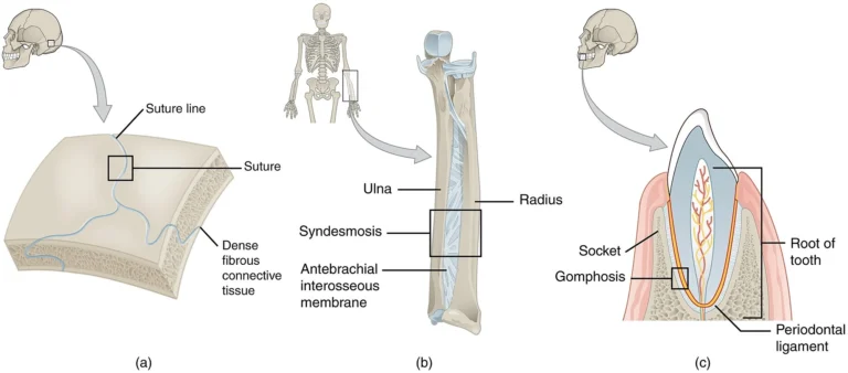
Sutures are immovable fibrous joints located exclusively between the bones of the skull.
They consist of interdigitating bony margins connected by short bundles of dense fibrous connective tissue, forming a highly stable articulation that maintains the integrity of the cranial vault.
Structure
Sutures are composed of thin layers of dense fibrous connective tissue with sharply interlocking bone edges. Collagen fibers anchor adjacent cranial bones, providing high tensile strength while permitting minimal elasticity during early development.
With age, these fibrous connections progressively ossify, leading to increased rigidity.
Types
Serrate sutures – saw-tooth interlocking pattern; provide maximal stability (e.g., sagittal suture)
Denticulate sutures – deep tooth-like interdigitations; enhance resistance to mechanical stress (e.g., lambdoid suture)
Plane sutures – relatively flat contacting surfaces; allow minimal movement (e.g., internasal suture)
Squamous sutures — overlapping beveled edges; permit slight gliding (e.g., temporoparietal suture)
Functional /Clinical
In infancy, sutures are separated by fontanelles, membranous gaps that facilitate skull deformation during childbirth and allow rapid brain growth. With maturation, sutures undergo synostosis, resulting in progressive fusion and reduced mobility.
Clinical Significance
Craniosynostosis – premature sutural fusion leading to abnormal skull morphology and potential impairment of brain development
Skull fractures – fracture lines may propagate along sutural planes due to the relative mechanical weakness of fibrous connections
Definition
A syndesmosis is a fibrous joint in which bones are connected by longer bundles of fibrous connective tissue, ligaments, or interosseous membranes.
Unlike sutures, syndesmoses permit a limited degree of movement depending on the length, elasticity, and orientation of the fibers.
Structure
The fibrous tissue connecting bones may form:
Ligaments – discrete bands of collagen fibers
Interosseous membranes – broad fibrous sheets connecting parallel bones
The collagen fibers are oriented to resist tensile stress and shear forces, providing both stability and controlled mobility.
Types
Distal tibiofibular syndesmosis
Connects tibia and fibula at the ankle
Stabilized by
anterior /posterior tibiofibular ligament
interosseous ligament
Interosseous membrane of the forearm
Connects radius and ulna
Distributes forces during forearm movements
Interosseous membrane of the leg
Connects tibia and fibula
Stabilizes the leg and transmits forces during weight bearing
Functional/Clinical
Syndesmoses play key biomechanical roles:
Stabilize long bones during movement
Transmit mechanical loads between adjacent bones
Maintain alignment of skeletal elements
Provide attachment sites for muscles
Clinical Significance
High ankle sprain
Injury to the distal tibiofibular syndesmosis resulting from excessive external rotation or dorsiflexion.
Such injuries compromise ankle stability and often require prolonged healing or surgical stabilization.
Definition
A gomphosis is a specialized fibrous joint in which a conical structure fits into a socket, forming a peg-and-socket articulation.
The only example in the human body is the articulation between a tooth and its alveolar socket.
Structure
The gomphosis consists of:
Tooth root
Alveolar bone of the maxilla or mandible
Periodontal ligament
The periodontal ligament is composed of dense collagen fibers that anchor the tooth to the
alveolar bone while allowing slight physiologic mobility.
Functional Role
Gomphoses serve several critical functions:
Secure teeth within alveolar sockets
Absorb mechanical forces generated during mastication
Distribute occlusal loads to surrounding bone
The slight mobility provided by the periodontal ligament prevents excessive stress on the tooth
and alveolar bone.
Clinical Relevance
Periodontal disease
Inflammation and degeneration of the periodontal ligament can weaken the gomphosis, leading to tooth mobility and eventual tooth loss.
Orthodontic tooth movement
Controlled mechanical forces stimulate bone remodeling within the periodontal ligament, allowing repositioning of teeth within the alveolar sockets.
Structural Stability
Fibrous joints provide maximal mechanical stability by firmly binding adjacent bones through dense collagenous connective tissue.
This rigidity is essential for maintaining precise anatomical alignment and preventing displacement under physiological and pathological loads, particularly in regions where movement would compromise structural integrity (e.g., cranial sutures).
Organs Protection
Fibrous articulations contribute directly to the formation of protective osseous compartments.
Cranial sutures interlock to create a continuous, mechanically resistant vault that safeguards the brain, ensuring that external forces are dissipated across a unified skeletal structure rather than transmitted to underlying neural tissue.
Force Transmission
Syndesmoses and interosseous membranes function as force-transmitting interfaces, redistributing mechanical loads between adjacent bones during dynamic activities such as locomotion.
This allows for load sharing, reduces peak stress on individual bones, and enhances overall biomechanical efficiency of the musculoskeletal system.
Adaptive Anchoring
Fibrous joints provide secure yet adaptable anchorage in systems requiring both fixation and micro-mobility.
The gomphosis exemplifies this, where the periodontal ligament allows minimal displacement to absorb and dissipate forcesduring mastication, preventing structural damage while maintaining positional stability.
Cranial Fusion
Premature fusion of cranial sutures disrupts normal skull expansion, leading to abnormal cranial morphology and potential increased intracranial pressure.
This condition highlights the critical balance between stability and growth in fibrous joints during development.
Syndesmotic Instability
Injuries to syndesmoses (e.g., distal tibiofibular joint) result in loss of interosseous tension and joint congruency, impairing force distribution across the limb.
Clinically, this leads to persistent instability, altered gait mechanics, and prolonged recovery compared to typical ligamentous injuries.
Anchorage Loss
Degeneration of the periodontal ligament in gomphoses leads to progressive loss of tooth stability, reflecting failure of fibrous anchoring mechanisms.
This results in tooth mobility, impaired mastication, and eventual tooth loss, demonstrating the functional importance of micro-mobility within rigid systems.
Joint Failure
Disruption of fibrous joints compromises load transmission pathways, leading to abnormal stress distribution, compensatory biomechanical changes, and increased risk of secondary injury.
Even minimal alterations in these “immobile” joints can produce significant functional deficits within the musculoskeletal system.